
Figure 8.
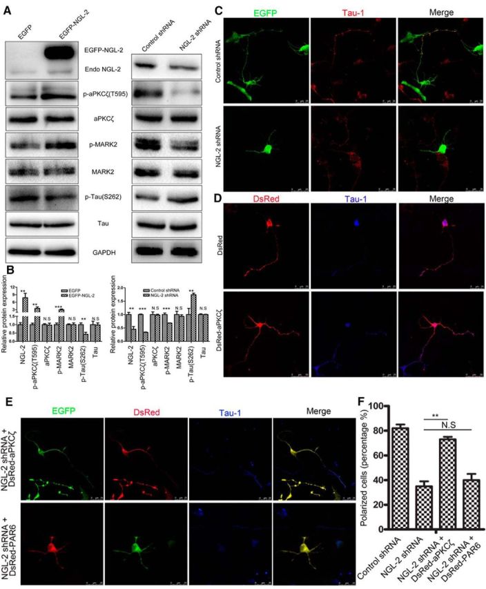
NGL-2 promotes axon differentiation by the aPKCζ/MARK2 signaling pathway. A, B, NGL-2 regulates tau phosphorylation by aPKCζ/MARK2 singaling pathway. A, Hippocampal neurons transfected by pEGFP-NGL-2 (left) or NGL-2 shRNA (right) were lysed and analyzed by Western blotting with indicated antibodies. Blotting with anti-GAPDH antibody showed equal amount of loading. B, The relative protein expression was normalized to GAPDH. The error bars represent the SD of three experiments. **p < 0.01, ***p < 0.001, Student's t test. N.S., Not significant. C–E, Overexpression of aPKCζ counteracts the polarity defect caused by NGL-2 suppression. Hippocampal neurons cotransfected with NGL-2 shRNA and DsRed-aPKCζ or DsRed-PAR6 were identified by GFP and DsRed. Axons were identified by anti-Tau1 antibodies. Scale bar, 25 μm. F, Quantification of the percentage of neurons that are polarized with a single axon [control shRNA, 82 ± 3% (n = 40); NGL-2 shRNA, 35 ± 4% (n = 40); NGL-2 shRNA + DsRed-aPKCζ, 73 ± 2% (n = 40); NGL-2 shRNA + DsRed-PAR6, 40 ± 5% (n = 42)]. The error bars represent the SD. *p < 0.001, Student's t test. N.S., Not significant.