Figure 5.
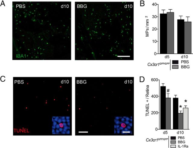
BBG and IL-1Ra inhibit subretinal inflammation-associated photoreceptor degeneration in Cx3cr1GFP/GFP mice. A, Representative accumulation of subretinal MP cells (choroidal/RPE flat mounts; green) at day 10 in PBS and BBG (25 mg/L) of Cx3cr1GFP/GFP mice treated by intravitreal (IVT) injections at days 3 and 7. Scale bar, 200 μm. B, MP density in the subretinal space of the light-challenge model of Cx3cr1GFP/GFP mice at days 5 and 10 treated with PBS and BBG at day 3 (animals kept until day 10 received a second IVT on day 7). (n = 6 per group, one-way ANOVA followed by Bonferroni's post-test, no differences were found between Cx3cr1GFP/GFP treated with PBS and BBG). C, Confocal microscopy of TUNEL-positive cells (red) through the ONL of a retinal flat mount at day 10 of the light-challenge model of Cx3cr1GFP/GFP mice treated with PBS or BBG (25 mg/L) by IVT injections. Scale bars: 50 μm, Inset, 10 μm. D, Quantification of the number of TUNEL-positive cell per retina of the light-challenge model of Cx3cr1GFP/GFP mice at days 5 and 10 treated with PBS, BBG (25 mg/ml), and IL-1Ra (150 mg/ml) at day 3 (animals kept until day 10 received a second IVT on day 7). (n = 5 per group, #p < 0.05; Mann–Whitney U test day 5 PBS vs day 5 BBG, *p < 0.05; one-way ANOVA followed by Dunnett's post-tests vs day 10 PBS).

