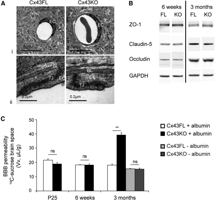
Figure 3.
BBB phenotype in the absence of astroglial Cx43. Ai, Representative images of the gliovascular unit ultrastructure in 3-month-old Cx43KO and Cx43FL (n = 6); Aii, details of TJs and BL. A, Astroglial perivascular endfeed; EC, endothelial cells. B, Western blot of the TJ proteins ZO-1, Claudin 5, and Occludin in 6-week-old and 3-month-old Cx43KO and Cx43FL cortex and hippocampus. GAPDH was used as the loading control (n = 3). C, BBB integrity in Cx43KO and Cx43FL mice at P25, 6 weeks, and 3 months of age was assessed by measuring the brain Vv (in microliters per gram) after in situ brain perfusion of [14C]sucrose under shear stress and increased hydrostatic vascular pressure [with (+) albumin, 180 mmHg, white and black bars] or under normal hydrostatic pressure and without shear stress [no (−) albumin, 120 mmHg, gray bars]. At P25 with albumin: Cx43FL, 21.6 ± 0.9 and Cx43KO, 19.0 ± 1.0, p = 0.7, n = 7; at 6 weeks with albumin: Cx43FL, 18.2 ± 0.2 and Cx43KO, 18.3 ± 0.8, p = 0.7, n = 7; at 3 months with albumin: Cx43FL, 18.1 ± 0.8 and Cx43KO, 39.3 ± 1.6, p = 0.005, n = 7; at 3 months without albumin: Cx43FL, 15.7 ± 0.2 and Cx43KO, 15.4 ± 0.3, p = 0.7, n = 7. Data are means ± SEMs. nsp > 0.05, **p < 0.001. Mann–Whitney two-tailed test.

