
Figure 7.
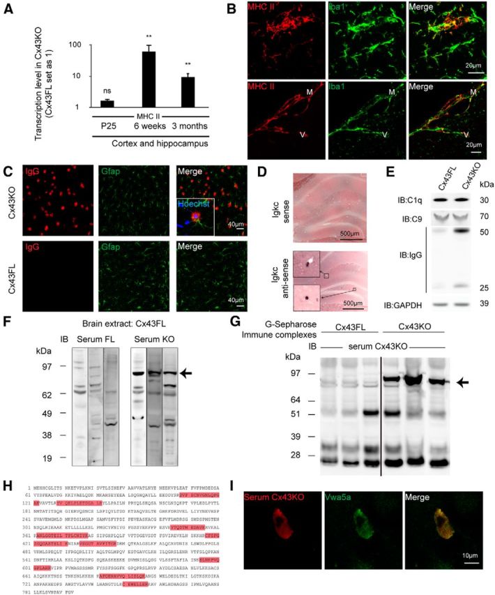
Humoral autoimmune response in the absence of astroglial Cx43. A, qPCR analysis of MHC II expression on Cx43KO and Cx43FL cortex and hippocampus at P25: 1.7 ± 0.2, p = 0.09, n = 4; at 6 weeks, 63.9 ± 38.0, p = 0.002, n = 4; at 3 months, 9.9 ± 2.7, p = 0.002, n = 5. Cx43FL values are set as 1. Data are presented as means ± SEMs. Mann–Whitney two-tailed test. nsp > 0.05, **p < 0.01. Cx43FL values are set as 1. B, Immunohistodetection of MHC II in the brain parenchyma and perivascular space of 6-week-old Cx43KO (n = 3). Microglia and macrophages are labeled with Iba1. MHC II labeling was absent in Cx43FL (our unpublished observation). C, Gfap (green) and IgG (red) double immunostaining in the cortex of 3-month-old Cx43KO and Cx43FL. Squared area shows a labeled astrocyte. D, Detection of Igkc transcript in Cx43KO hippocampus by ISH, with detail of labeled B cells (squared area). Sense probe is the negative control (n = 3). E, C1q, C9, and IgG heavy and light chain immunodetection on Western blots of 3-month-old Cx43KO and Cx43FL brain proteins (n = 3). F, Immunoblot (IB) of Cx43FL brain proteins using three different Cx43KO or Cx43FL sera. A band around 90 kDa is selectively revealed by Cx43KO sera (arrow; n = 7). G, G-Sepharose-purified immune complexes from Cx43KO and Cx43FL brain immunoblotted with a Cx43KO serum. A band around 90 kDa is selectively revealed in Cx43KO-purified proteins (arrow; n = 12). H, Matrix-assisted laser desorption/ionization time-of-flight analysis of these immune complexes revealed the presence of nine peptides (in red) belonging to Vwa5a in Cx43KO immune complexes only (n = 6). I, A Vwa5a-transfected primary astrocyte immunolabeled by both a Cx43KO serum (red) and a Vwa5a-specific antibody (green; n = 3).