Figure 2.
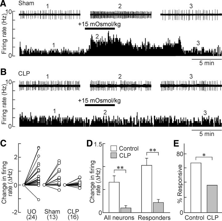
Osmosensitivity of OVLT neurons is lost in CLP rats. A, Ratemeter records (5 s bin width) showing the firing rate of a single OVLT neuron in an explant prepared from a sham rat. Note the reversible excitatory effect of the hyperosmotic stimulus (bar). Traces above the ratemeter plot show excerpts of the raw single-unit activity recorded at the time points indicated by the numbers. B, Response of an OVLT neuron recorded from a CLP rat (layout as in A). C, Two-point plots show the changes in firing rate induced by hyperosmotic stimulation in all of the OVLT neurons tested in unoperated (UO), sham and CLP rats (UO 24 neurons/9 rats; sham, 13 neurons/7 rats; CLP, 16 neurons/5 rats). D, Bar graphs show mean ± SEM changes in firing rate induced by hypertonicity in all of the OVLT neurons tested in control rats (UO + sham; 37 neurons/16 rats) and CLP (16 neurons/5 rats) or in the specific subset of OVLT neurons that showed at least a 15% increase in firing rate (responders: control, 25 neurons/16 rats; CLP, 6 neurons/5 rats). E, Bar graphs show proportions of responders in controls (25 neurons/16 rats) and CLP animals (6 neurons/5 rats). *p < 0.05; **p < 0.01.

