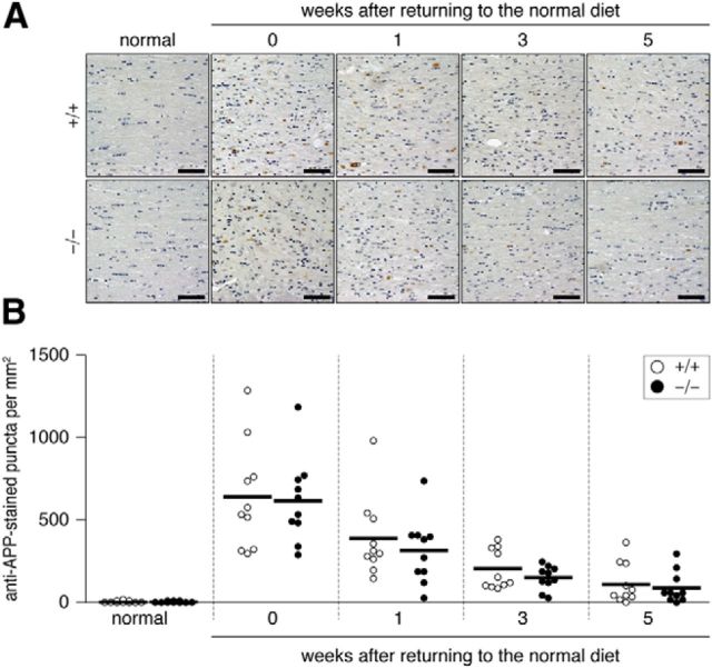
Figure 6.
A, Axonal damage induced in the dorsal corpus callosum by the cuprizone treatment. Stained images of damaged axons are shown. Brain sections from wild-type (+/+) and Ptprz-deficient (−/−) mice were stained with a specific antibody against APP (anti-APP) and then stained with hematoxylin. Scale bars, 50 μm. B, Scatterplot of the anti-APP-stained puncta in the dorsal corpus callosum. Open circles, Wild-type mouse; closed circles, Ptprz-deficient mouse. n = 10 for each group. No genotypic differences were detected by two-way ANOVA.

