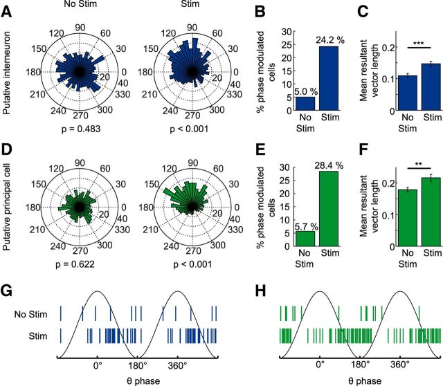
Figure 8.
Stimulation of cholinergic MSDB neurons increased spike-phase coupling in area CA3 of the dorsal hippocampus. A, D, Spike phase distribution of a single putative interneuron (A) or principal cell (D) during the baseline (No Stim, left) or stimulation (Stim, right) period. B, E, The number of putative interneurons (B) and putative principal cells (E) that fired significantly phase coupled (p < 0.05, Kuiper's test) to the local theta rhythm increased during optogenetic stimulation from 6–29 (from a total of 120 putative interneurons recorded from 22 mice) and from 10–50 (from a total of 176 putative principal cells recorded from 24 mice), respectively. Spike numbers of baseline and stimulation conditions were matched by random spike deletion. C, F, The mean resultant vector length averaged across putative interneurons (C) and putative principal cells (F) increased significantly with respect to baseline during the stimulation periods (bar graphs show mean ± SEM, **p < 0.01, ***p < 0.001, Wilcoxon signed-rank test). G, H, Mean resultant vector angles of significantly theta-phase-locked putative interneurons (G) and putative principal cells (H). Note that spiking of putative interneurons clusters around the descending phase, whereas spiking of putative principal cells clusters around the trough of the local theta rhythm.

