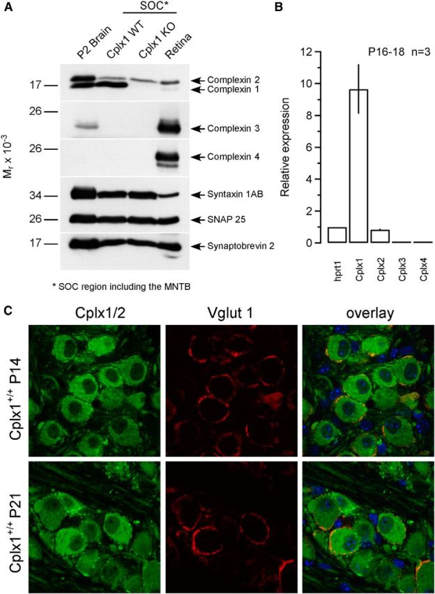
Figure 1.

Analysis of Cplx expression in the mouse MNTB. A, P2 fraction from adult brain and tissue homogenates from adult retina and the MNTB region of the superior olivary complex (SOC) cut out from acute brainstem slices of P17 Cplx1+/+ and Cplx1−/− littermates (10 μg per lane) were analyzed by SDS-PAGE using antibodies against Cplx1/2, Cplx3, and Cplx4, as well as antibodies against the presynaptic marker proteins Syntaxin 1AB, Synaptobrevin 2, and SNAP 25. B, Validation of mRNA expression level of different Cplx isoforms in the AVCN region cut out from acute brainstem slices of P16–P18 Cplx1+/+ mice by quantitative real-time PCR. The expression level was calculated according to 2−(Ctgene − Cthprt1), where Ctgene and Cthprt1 represent the threshold levels of detection for the genes tested and for the housekeeping gene, respectively. The relative expression was then obtained by normalizing the expression levels to that of the housekeeping gene hprt1 (n = 3; technical replicates). C, Cplx1/2 localization patterns in the developing mouse calyx of Held. Immunofluorescence images representing projections of confocal sections of MNTB Cplx1+/+ mice costained with an anti-Cplx1/2 antibody (green, left), and an anti-Vglut1 antibody (red, middle) at P14 and P21. Cplx1/2 immunofluorescent MNTB PNs are surrounded by ring-like Cplx1/2- and Vglut1-positive structures representing calyx terminals. Right, The corresponding overlay images.
