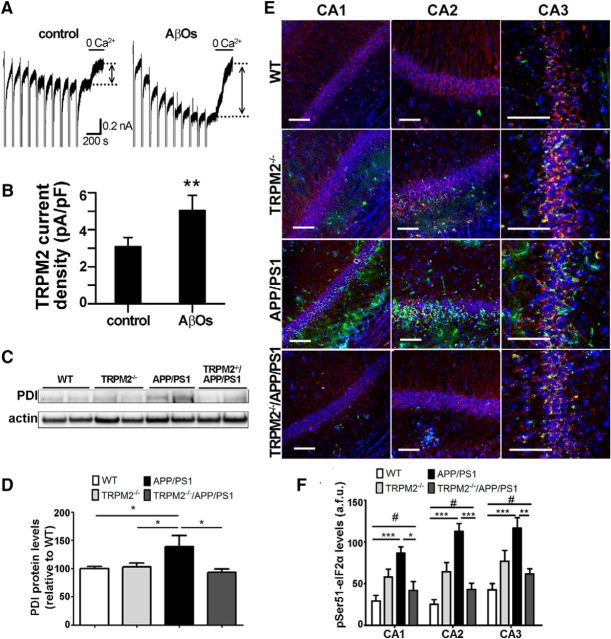
Figure 1.
TRPM2 is responsive to AβOs and contributes to increased ER stress in APP/PS1 mice. A, Representative traces show that NMDA facilitated ADPR-induced TRPM2 currents evoked in cultured hippocampal neurons from CD1 mice in the absence or presence of AβOs. These could be inhibited by omitting Ca2+ from the extracellular recording solution. TRPM2 current amplitude was determined as indicated by bidirectional arrows positioned between the stippled lines. B, Summary bar graphs show that treatment with 1 μm AβOs for 24 h increases TRPM2 current amplitude and density. Data from at least five cells for each treatment were analyzed with Student's t test. **p < 0.01. C, D, Immunoblot analysis of PDI levels in hippocampal extracts. At least four brain extracts obtained from each genotype were analyzed. E, F, Immunofluorescence analysis of phospho-eIF2α(Ser51) (green) levels in CA1, CA2, and CA3 areas of mouse hippocampus. βIII-tubulin (red) and Hoechst dye (blue) were used to label neurons and nuclei, respectively. At least four coronal slices from each mouse brain and at least three brains of each genotype were used for immunostaining. *p < 0.05 (one-way ANOVA followed by Tukey's post hoc test). **p < 0.01 (one-way ANOVA followed by Tukey's post hoc test). ***p < 0.001 (one-way ANOVA followed by Tukey's post hoc test). #p > 0.05 when comparing pSER51 WT levels to TRPM2−/−APP/PS1 levels. Scale bar, 90 μm.

