
Figure 4.
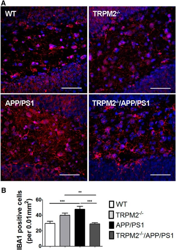
Decreased microglial activation in brains of TRPM2−/−/APP/PS1 mice. Levels of activated microglia in DG (A, B) area of mouse hippocampus were analyzed by Iba1 immunostaining (red). Hoechst dye (blue) was used to label nuclei. At least four coronal slices from each mouse brain and at least 3 brains of each genotype were used for immunostaining. **p < 0.01 (one-way ANOVA followed by Tukey's post hoc test). ***p < 0.001 (one-way ANOVA followed by Tukey's post hoc test). Scale bar, 90 μm.