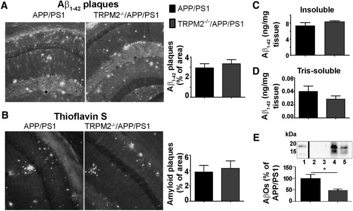
Figure 5.
Normal plaque load in brains of TRPM2−/−/APP/PS1 mice. Aβ levels in mouse hippocampus. A, B, Brain slices were stained for plaques with anti-Aβ(1–42) IgG (A) and thioflavin S (B). At least four coronal slices from each mouse brain and at least three brains of APP/PS1 and TRPM2−/−/APP/PS1 mice were used for each immunostaining experiment. C, D, Levels of Aβ(1–42) of insoluble (C) and Tris-soluble (D) fractions of at least 6 hippocampi of APP/PS1 and TRPM2−/−/APP/PS1 mice were analyzed by ELISA. E, Western blot analysis of intracellular and cell membrane-bound fractions of hippocampus. The lower band corresponds to the trimers, and the upper band to the tetramers of Aβ(1–42). At least five extracts obtained from each genotype were analyzed. *p < 0.05 (Student's t test).

