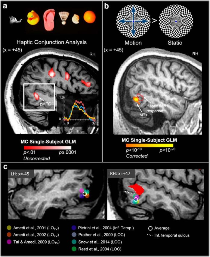
Figure 8.

The tactile response in pMTG in patient M.C. lies dorsal to MT+, LOTV, and LOC foci identified in previous fMRI studies of haptic object perception. a, In the haptic conjunction fMRI analysis, we observed a large cluster of activation at the posterior end of the medial temporal gyrus (pMTG) in the RH of patient M.C. The inset (bottom right) shows the time course of activation in the pMTG cluster. b, Although M.C. cannot identify stationary objects using vision, she can nevertheless detect motion, a condition known as Riddoch's phenomenon. Accordingly, M.C. showed strong activation in area MT+ when she viewed moving versus static checkerboard displays. The haptic shape-related area in pMTG (dotted red line) lies dorsal and anterior to MT+. c, Sagittal slices showing the left and right temporal lobes of M.C.'s brain. The haptic response in right pMTG in M.C. is shown in red (p = 0.01 uncorrected). Filled circles (see color key) show the position in Talairach y- and z-coordinates of foci of visuohaptic responses in LOTV, a putative subregion of LOC that responds to both visual and tactile stimuli (as identified by Amedi et al., 2001, 2002, 2009) and responses in LOC and inferior temporal cortex (as identified in previous fMRI studies involving haptic perception of 3D solid objects). Sagittal slices are shown from the average position of peak activation across all studies (LH: x = −45, RH: +47). The average position of peak haptic responses in x-, y-, and z-coordinates across all studies is represented by the open (white) circle. The inferior temporal sulcus (dotted white line) marks the boundary between inferior and middle temporal gyri.
