Figure 6.

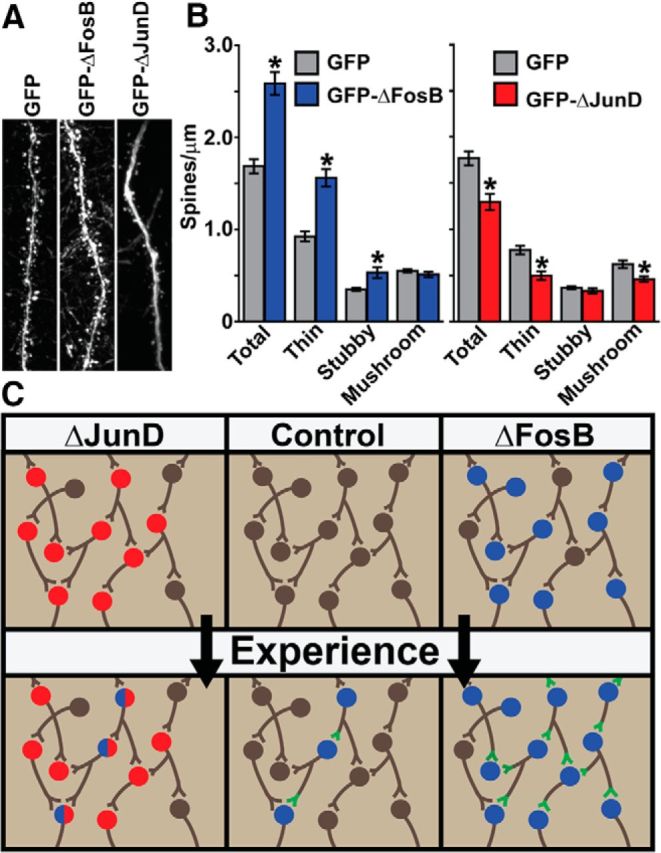
ΔFosB regulates hippocampal spine formation. Mice received hippocampal infusions of HSV–GFP, HSV–GFP–ΔFosB, or HSV–GFP–ΔJunD and dendritic spine analysis was conducted on hippocampal CA1 pyramidal neurons. A, Representative dendrites showing spines from GFP+ hippocampal CA1 neurons. B, Quantitation of spines in CA1 neurons shows ΔFosB (blue; n = 30) had significantly more total spines, thin spines, and stubby spines compared with GFP alone (gray; n = 30; *p < 0.001), with no significant difference in mushroom spines. ΔJunD (red; n = 19) had significantly fewer total spines, thin spines, and mushroom spines compared with GFP alone (gray; n = 22; *p < 0.001), with no significant difference in stubby spines. Error bars indicate mean ± SEM. C, Model depicting experience-dependent changes in hippocampal cells under control conditions or when virally transduced with ΔJunD (red) or ΔFosB (blue). Green indicates changes in synaptic structure/function resulting from ΔFosB expression within a circuit that may underlie learning.
