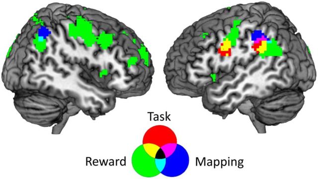
Figure 4.
Overlap of all three MVPAs. Regions informative about the mapping during delay 1 are shown in red, regions informative about the task during task execution are shown in blue, and regions informative about the reward during task execution are shown in green. The informative regions of all three analyses overlap in the left inferior parietal cortex, whereas regions informative about mappings and rewards overlap in the right inferior parietal cortex. This shows that brain areas that represent an association between tasks and rewards early in the trial represent actual tasks and rewards or their effects at a later stage in the trial.

