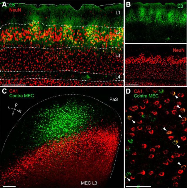
Figure 1.
Layer 3 of the MEC: homogeneous layout and organization of long-range projections. A, Superimposed staining for calbindin (green) and NeuN (red) showing the homogeneous and uniform distribution of neuronal somata in layer 3 of the MEC. Note comparison of the modular organization of the adjacent layer 2. Scale bar, 250 μm. B, Parasagittal section through the MEC stained for calbindin (top, green) and NeuN (bottom, red) from A. Scale bar, 250 μm. C, Tangential section through MEC layer 3 showing retrogradely labeled neuronal somata following injection of CTB-Alexa 488 (green) and CTB-Alexa 546 (red) in contralateral MEC (contra MEC) and ipsilateral hippocampus (CA1), respectively. The border of the MEC is outlined (dotted line). PaS, Parasubiculum. Scale bar, 250 μm. D, High-magnification of a parasagittal section from a dual-retrograde neuronal tracing experiment as in C. Arrowheads indicate double-labeled neurons. Labels as in C. Scale bar, 100 μm. D, Dorsal; L, lateral; M, medial; V, ventral.

