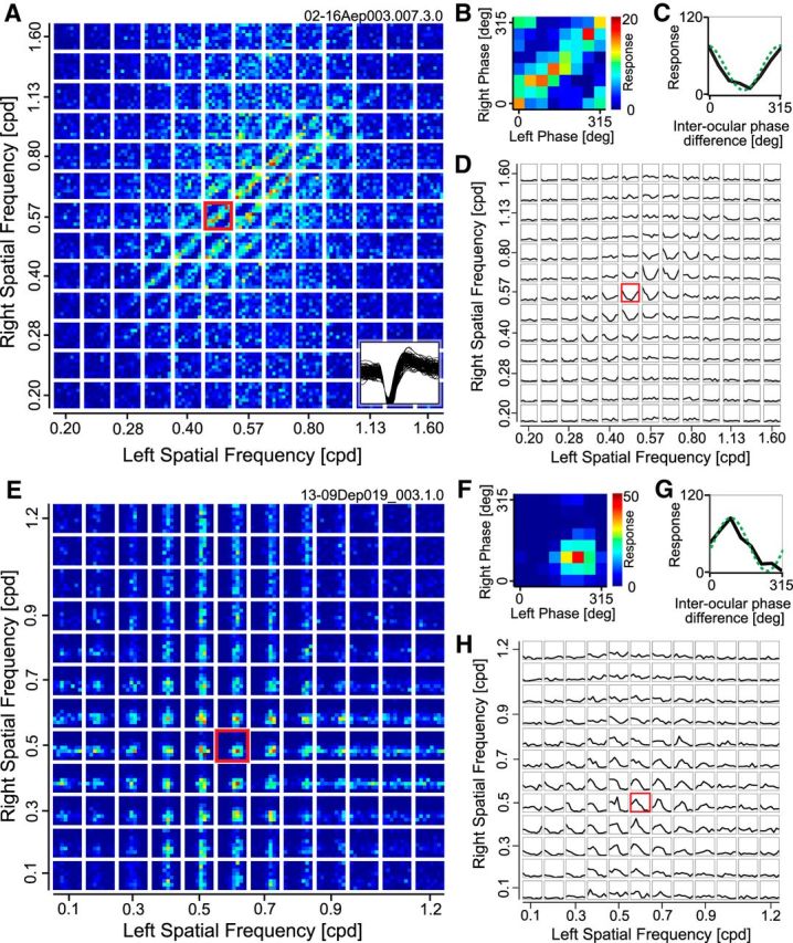
Figure 4.

A–D, Responses are shown for a representative complex cell to stimuli containing various combinations of phases and SFs between the two eyes. A, The entire data are response strengths in a 4-D parameter space (sfL, sfR, phL, phR), and presented here as multiple cross sections (phL, phR) at each (sfL, sfR) for clarity. Each small domain is a binocular phase-selectivity map (phL, phR), illustrating responses to various combinations of left and right phases. These small maps are arranged as a 13 × 13 matrix of left and right SFs (sfL, sfR). Color scale shows the number of spikes collected for each pair of phases for the two eyes. One of the phase selectivity maps is marked with a red border and is magnified in B. Example spike waveforms of this cell are drawn superimposed at the bottom-right corner (100 spikes). B, Selectivity to binocular phase combination for the highlighted SF pair in A is shown. Strong responses are observed along a 45° diagonal where the interocular phase difference was constant. C, One-dimensional tuning curve to interocular phase difference was calculated from a binocular phase selectivity map shown in B by integrating responses to the same interocular phase difference between the two eyes. A sinusoid was fitted to the tuning curve to determine the modulation amplitude (green broken curve). D, Tuning curves to interocular phase difference are represented for all binocular combinations of SFs. The highlighted SF pair shown in C is indicated with a red border. The optimal SF and orientation for the dominant eye of this cell were 0.64 cpd and 19°, respectively. Values were similar for the nondominant eye (0.61 cpd, 8°). E–H, Responses are shown for a representative simple cell to stimuli containing various combinations of phases and SFs between the two eyes. E–H, The same format as A–D. Optimal SF and orientation for the dominant eye of this cell were 0.56 cpd and 9°, respectively. Values were similar for the nondominant eye (0.53 cpd, 30°).
