
Figure 10.
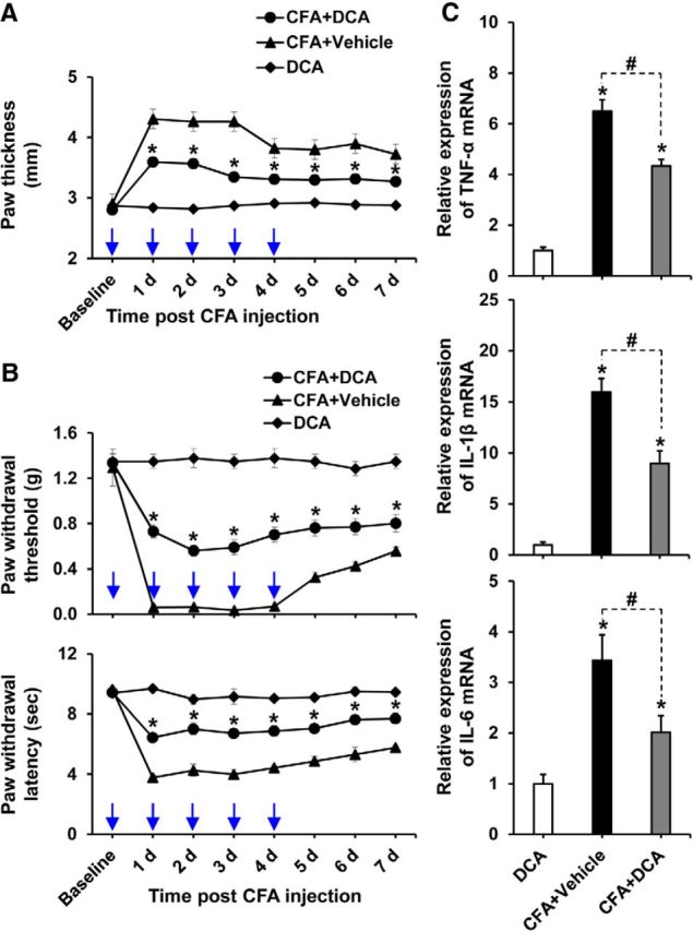
Pharmacological inhibition of PDKs attenuated CFA-induced paw edema, pain behaviors, and expression of proinflammatory cytokines. A, B, To examine the role of PDKs, DCA (10 mg/kg body weight, 10 μl) or vehicle (saline, 10 μl) was administered daily to the CFA-injected hindpaws. A group of animals received only DCA in left hindpaws. CFA-induced increase in paw thickness (A), PWT to force, and PWL to heat (B) was assessed up to 7 d after CFA injection. C, Hindpaw tissues were collected at day 3 after CFA administration from vehicle or DCA-treated animals to assess the expression of proinflammatory cytokines. The relative mRNA expression of TNF-α, IL-1β, and IL-6 in the hindpaw tissues was evaluated by real-time RT-PCR. Results for mRNA expression are displayed as the fold increase of gene expression normalized to GAPDH. *p < 0.05, between CFA+DCA and CFA+Vehicle groups, or versus the control animals. #p < 0.05 between indicated groups (Student's t test, Mann–Whitney test for PWT). A, B, n = 6; C, n = 3. Data are mean ± SEM. A, B, Arrows indicate the time points of DCA or vehicle administration.