
Figure 11.
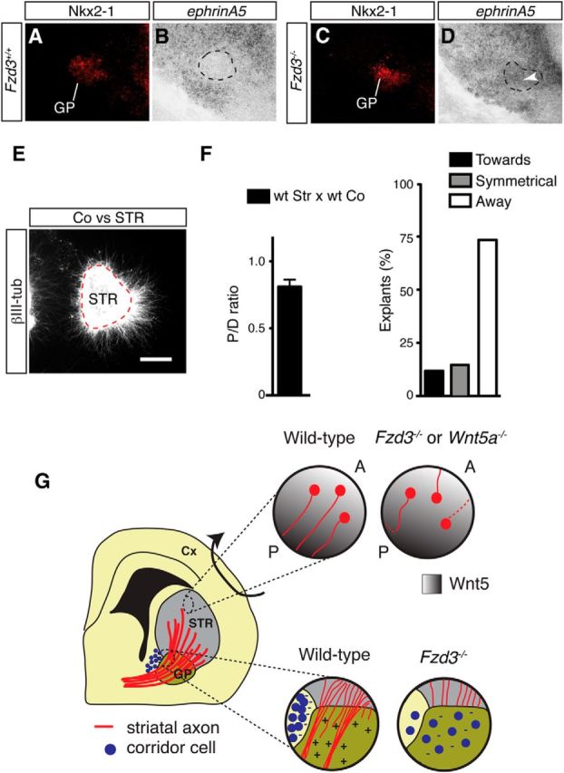
Corridor cells induce striatal axon repulsion. A–D, Double staining combining immunohistochemistry for Nkx2-1 (A, C) and in situ hybridization for ephrinA5 (B, D) in coronal sections from E13.5 embryos. EphrinA5-positive cells are present in the GP of Fzd3−/− but not wild-type embryos (indicated by arrowhead). Number of mice analyzed: E13.5 Fzd3+/+, n = 3; E13.5 Fzd3−/−, n = 3. E, Collagen matrix cocultures of E14.5 striatal (STR) and corridor cell (Co) explants maintained for 2 DIV and immunostained for βIII-tubulin. Coexplants are at the left. Dotted line indicates STR explant. F, Left graph, Quantification of the P/D ratios of STR explants in cocultures as in E. P/D < 1.0 indicates repulsion. n = 34 cocultures. Right graph, Quantification of the number of STR explants that show symmetrical axon growth or growth toward or away from coexplants. Scale bars: A–D, 220 μm; E, 160 μm. G, Schematic representation of a coronal section of the embryonic mouse brain showing the striatum (STR) and GP. In wild-type embryos, MSNs in the striatum send their axons posteriorly. These axons project into (striatopallidal pathway) or through (striatonigral pathway) the GP. An anterior (A) low, posterior (P) high Wnt5a/Wnt5b gradient is present in the striatum, and MSN axons are attracted by Wnt5s in vitro. Fzd3−/−, and Wnt5a−/− mice display aberrant caudal, lateral, and medial MSN projections. These data support a model in which Wnt5s guide Fzd3-postive MSN axons along the AP axis of the STR. In Fzd3−/− mice, all striatal axons accumulate at the GP and do not enter this structure. Corridor cells express axon repellents for MSN axons and are mislocalized in the GP of Fzd3−/− mice. The normally attractive effect of the GP on MSN axons is lost in the absence of Fzd3. This suggests that patterning of corridor cells by Fzd3 is required to create permissive corridors for MSN axon growth. Cx, Cortex.