


Figure 2.
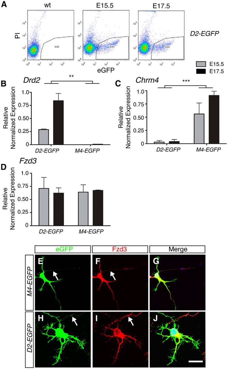
Molecular profiling of Frizzled expression in striatopallidal and striatonigral MSNs. A, Scatter plots showing the distribution of EGFP-positive MSNs purified from E15.5 and E17.5 D2-EGFP embryos compared with reference tissue. EGFP-positive neurons (outlined area) were selected by FACS. PI, Propidium iodide; wt, wild-type. B–D, Quantitative PCR was used to determine relative expression of Dopamine receptor D2 (Drd2; marks striatopallidal MSNs), Cholinergic receptor, muscarinic 4 (Chrm4; marks striatonigral MSNs), and Frizzled3 in D2-EGFP+ or M4-EGFP+ cells obtained by FACS at E15.5 or E17.5. Drd2 is expressed in D2-EGFP+ but not in M4-EGFP+ cells, whereas Chrm4 is expressed in M4-EGFP+ but not D2-EGFP+ cells, confirming the specificity of the FACS procedure. Of 10 Frizzleds tested, only Fzd2 (data not shown) and Fzd3 showed detectable expression in E15.5 and E17.5 MSNs. n = 3 independent experiments. **p < 0.01 (two-way ANOVA). ***p < 0.001 (two-way ANOVA). E–J, Coimmunostaining for EGFP and Frizzled3 in dissociated striatal neuron cultures generated from E15.5 M4-EGFP or D2-EGFP mice analyzed at 3 DIV. Arrows indicate axons. M4-EGFP n = 3 litters, D2-EGFP n = 3 litters. Scale bars: E–J, 40 μm.