
Figure 7.
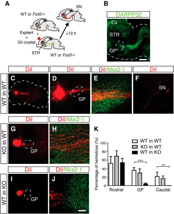
Frizzled3 non–cell-autonomously regulates the entry of striatal axons into the GP. A, Schematic illustrating the transplantation of Fzd3+/+ (WT) or Fzd3−/− explants into WT or Fzd3−/− hemislices followed by striatal pathway analysis using DiI labeling. STR, Striatum. B, E14.5 hemislice cultured for 3 DIV and subjected to whole-mount immunostaining for DARPP32 (green). Cx, Cortex. Dotted lines indicate boundaries of hemislice and GP. C–J, Examples of WT and Fzd3−/− explants transplanted into hemislices and analyzed at DIV3. DiI labeling in red and immunostaining for Nkx2-1, to label the GP, in green. C, Dotted line outlines hemislice. D, G, I, Dotted line outlines the GP. DiI-labeled striatal axons from WT explants fail to enter the GP in KO hemislices (I, J). K, Quantification of the number of hemislices showing DiI-labeled projections from transplanted explants rostral to the GP, in the GP, or caudal to the GP. **p < 0.01 (Kruskal–Wallis test). ***p < 0.001 (Kruskal–Wallis test). Data represent mean ± SEM. Scale bars: B, 100 μm; C, 2.2 mm; D, G, I, 75 μm; E, F, H, J, 20 μm.