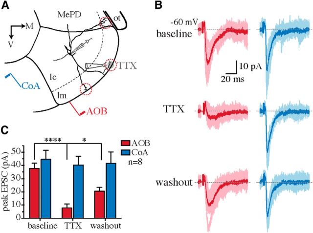
Figure 4.
Focal blockade of synaptic transmission at distal dendrites disrupts AOB but not CoA inputs. A, Schematic of focal pressure application of TTX at distal dendritic branches (red circles) of MePV principal neurons. MePD, Posterodorsal medial amygdala; ot, optic tract; lm, molecular layer; lc, cellular layer. B, AOB (red) and CoA (blue) EPSCs (Vhold = −60 mV) recorded before (top) and after (middle) puffing 1 μm TTX at distal dendrites and following washout (bottom). Whereas AOB EPSCs were blocked by TTX (left), CoA input remained intact (right). C, Summary data showing the effect of focal TTX application on the peak amplitude of evoked EPSCs (mean ± SEM; n = 8 cells). *p < 0.05; ****p < 0.0001 (two-way ANOVA with Bonferroni's test).

