
Figure 5.
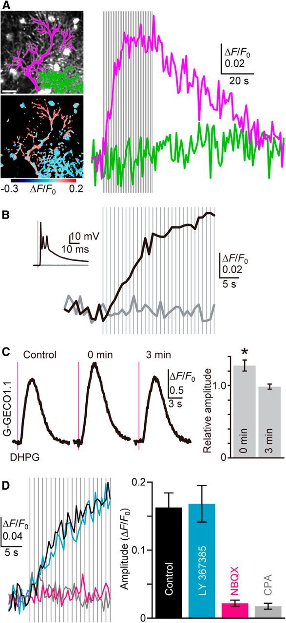
CF activity-induced ER Ca2+ overloading. A, Representative time courses of mean ΔF/F0 throughout the dendrite of a stimulated PC (magenta in the top image, magenta trace) and a nonstimulated neighboring PC (green in the top image, green trace) upon repetitive CF inputs (30 stimuli at 1 Hz, gray vertical lines) indicate CF-induced ER Ca2+ dynamics in PCs. The pseudo-color image shows the average of 20 consecutive frames during CF stimulation. Scale bar, 20 μm. B, CF stimulation (1 Hz, gray vertical lines) just above the threshold induced EPSP (black inset trace) and G-CEPIA1er response (black trace). In contrast, no EPSP (gray inset trace) and G-CEPIA1er responses (gray trace) were observed in the same PC upon stimulation just below the threshold. C, Cytoplasmic Ca2+ measurement with G-GECO1.1. Ca2+ mobilization was induced by pressure ejection of DHPG (1 mm, 50 ms, 10–30 psi, magenta vertical line), either 5–10 s (0 min) or 3 min after CF stimulation (30 pulses at 1 Hz). Representative time courses of ΔF/F0 indicate the transient enhancement of mGluR1-induced Ca2+ mobilization after CF stimulation. The summarized bar graph for the amplitude of the responses normalized by the amplitude of the control is shown. n = 7 (mean ± SEM). *p = 0.00559, t test. D, Pharmacological characterization of CF-induced ER Ca2+ dynamics. G-CEPIA1er responses upon CF inputs (1 Hz, gray vertical lines) in the control condition (black), or in the presence of LY367385 (100 μm, cyan), NBQX (10 μm, magenta), or CPA (50 μm, gray) are shown. Amplitude is defined as the maximum increase in ΔF/F0 during 20 pulses of CF stimulation. n = 6–13 (mean ± SEM).