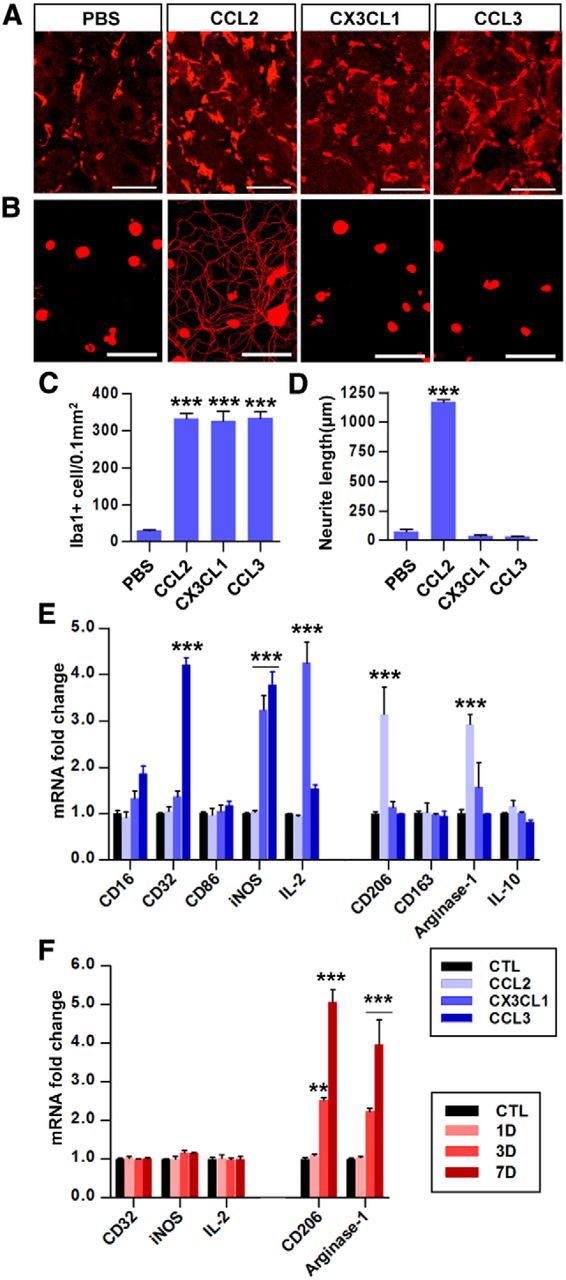
Figure 6.

CCL2 is sufficient for enhancement of axon regenerative capacity by mobilizing M2-like macrophages. A, Representative images of Iba1 staining in L5 DRG sections obtained 7 d after intraganglionic injection of PBS or CCL2, CXCL1, and CCL3. Scale bars, 50 μm. B, Representative images of neurons cultured from L5 DRGs dissected from animals with intraganglionic injection of PBS, or CCL2, CXCL1, and CCL3. Scale bars, 100 μm. C, Quantitative comparison of the number of Iba1-positive macrophages in DRGs. N = 3 animals per group. ***p < 0.001. D, Quantitation of neurite outgrowth. The culture period was 15 h and DRG neurons and their neurites were visualized by immunofluorescence staining for β III tubulin. N = 3–4 animals per group. ***p < 0.001. E, Real-time RT-PCR results for M1 and M2 markers in cultured macrophages treated with CCL2, CX3CL1, or CCL3 for 24 h. N = 3 independent cultures per group. ***p < 0.001 compared with the control (untreated) condition. F, Real-time RT-PCR results for of M1 and M2 marker gene expression in MACS-separated (using CD68 antibody) macrophages from the L4 and L5 DRGs at the indicated time points after SNI (CD68-positive fraction). N = 4 animals per group. **p < 0.01 and ***p < 0.001 compared with control values, respectively, by one-way ANOVA followed by Tukey's post hoc analysis.
