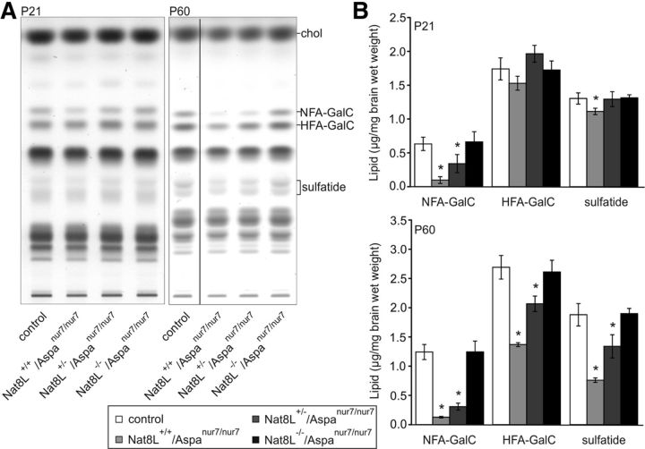
Figure 12.
Myelin sphingolipid levels are normalized in Aspanur7/nur7 mice with an additional Nat8L deficiency. A, Lipids were isolated from total brain of Nat8L+/+/Aspa+/+ or Nat8L+/+/Aspanur7/+ (control), Nat8L+/+/Aspanur7/nur7, Nat8L+/−/Aspanur7/nur7, and Nat8L−/−/Aspanur7/nur7 mice at the age of P21 and P60. Total lipids were separated by HPTLC and quantified by densitometry. The amount of lipids corresponding to 1 mg wet weight of brain tissue was applied per lane. Positions of standard lipids run on the same TLC plate are indicated (chol, cholesterol; NFA-GalC, nonhydroxy fatty acid-containing galactosylceramide; HFA-GalC, 2-hydroxy fatty acid-containing galactosylceramide). All samples from P60 mice were separated on the same TLC plate, however, several nonrelevant lanes between the first and second lane shown in the figure were omitted and a merged image is shown. B, Quantitative analysis of the lipids demonstrate a statistically significant reduction in NFA-GalC in Nat8L+/+/Aspanur7/nur7 and Nat8L+/−/Aspanur7/nur7 mice, but not in Nat8L−/−/Aspanur7/nur7 mice, compared with control mice. At P21, sulfatide was only reduced in Nat8L+/+/Aspanur7/nur7 mice. At P60, all quantified sphingolipids were significantly reduced in both, Nat8L+/+/Aspanur7/nur7 and Nat8L+/−/Aspanur7/nur7 mice compared with control littermates, demonstrating demyelination and the worsened phenotype from P21 to P60. There was no statistically significant difference between Nat8L−/−/Aspanur7/nur7 mice and control mice (*p < 0.05, t test, n = 3–4).

