
Figure 6.
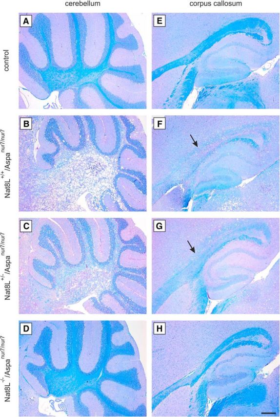
Luxol Fast Blue staining. A–H, Luxol Fast Blue staining of sagittal paraffin embedded brain sections of 60-d-old animals. Spongy degeneration and vacuoles formation go along with demyelination in Nat8L+/+/Aspanur7/nur7 (B, F) mice. Demyelination is demonstrated by less intense Luxol Fast Blue staining (arrows in F and G) shown, for example, in the white matter of cerebellum (A–D) and corpus callosum (E–H) compared with control littermates (A, E). Nat8L−/−/Aspanur7/nur7 (D, H) did not show any indication of myelin loss compared with controls. Scale bar, 200 μm.