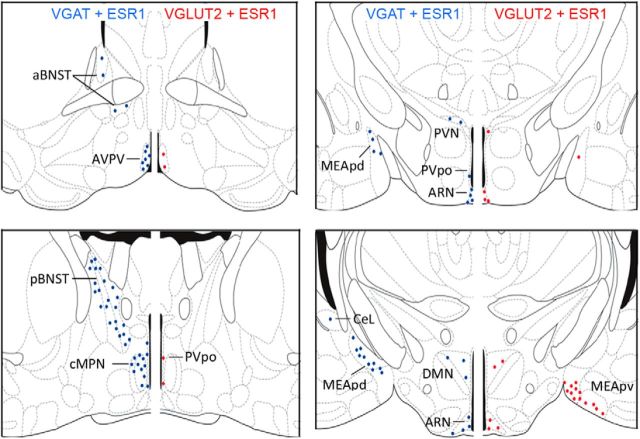
Figure 2.
Distribution and density of ESR1-expressing GABA and VGLUT2 neurons in the female mouse brain. Schematic diagrams adapted from Franklin and Paxinos (1997) showing the topography of VGAT-ESR1 (blue) and VGLUT2-ESR1 (red) neurons at four different levels through the female mouse forebrain. Each dot represents 10 dual-labeled cells in a 30-μm-thick coronal brain section. aBNST, anterior bed nucleus of the stria terminalis; CeL, lateral central amygdala; DMN, dorsomedial nucleus; MEApd, posterior dorsal medial amygdala; MEApv, posterior ventral medial amygdala; pBNST, posterior bed nucleus of the stria terminalis; PVpo, preoptic periventricular nucleus; PVN, paraventricular nucleus.

