Figure 2.

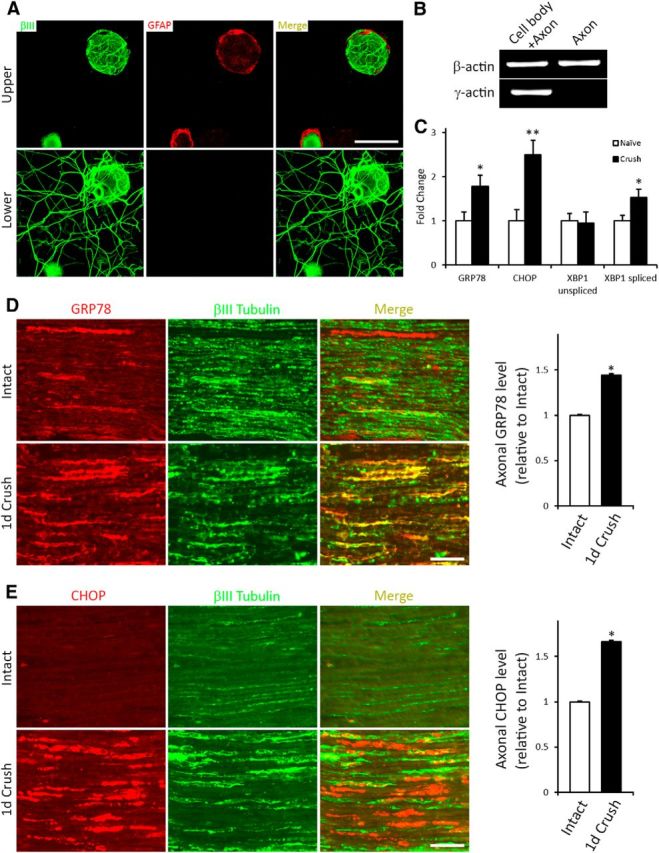
The UPR is elevated in injured axons. A, Neuronal DRG cultures grown on insert membranes and processed to detect the neuron/axon marker βIII tubulin (green) and the Schwann cell/satellite glial cell marker GFAP (red) immunofluorescence as indicated, with focal plane of images taken being either above (top) or below (bottom) the membrane. The presence of βIII tubulin and the absence of GFAP signal in the lower membrane indicate that only axons extend through the pores to grow on the underside of the membrane. Scale bar, 50 μm. B, qRT-PCR of RNA from “axon only” or “cell body + proximal axon” preparations. The absence of γ-actin confirms purity of “axon only” extract. C, qRT-PCR analysis of axonal mRNA levels of UPR-related genes relative to naive controls from 1 d crush injury-conditioned DRG neurons assayed 48 h in vitro on transwell inserts (N = 4 experimental replicates). *p < 0.01. **p < 0.01. Representative sections of sciatic nerve immediately proximal to the crush injury site or from intact control sciatic nerve processed to detect expression of the UPR proteins GRP78 (D) or CHOP (E) and colocalized with the axonal marker β-III tubulin. Histograms (right) summarize alterations in expression of the markers in the 1 d injured axons (1 d Crush) relative to Intact control. Scale bar, 100 μm. *p < 0.01. N = 3 animals and 37–44 fields analyzed/condition. The expression of both UPR-associated proteins is significantly elevated in the 1 d injured axons, with increased extra axonal expression also evident for CHOP.
