
Figure 2.
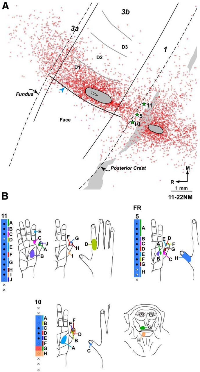
A, Composite plot of labeled neurons following injection of FR in D1 representation of normal Monkey 11-22NM shown on drawing of a section of the flattened cortex. Dashed lines indicate fundus and posterior crest of the central sulcus. Solid lines indicate approximate locations of the rostral and caudal borders of area 3b. Gray ovals outline the region of injection core. Open circles represent FR-labeled neurons. There are only few labeled neurons in the face representation lateral to HFS (blue arrowhead). HFS and the border between digits (gray curved lines) were located from myelin-stained sections. Gray shaded area on the right represents the region that was damaged in few of the sections during tissue processing. Stars indicate approximate locations of the electrode penetration sites for which receptive fields are shown in B. B, Receptive fields of neurons at and around the FR injection site shown for three adjacent electrode penetration sites. Injection of FR was centered at penetration 5. Receptive fields and responses of neurons at the recording sites marked by uppercase letters are shown by corresponding letters on adjacent figurines of the hand and face. Other conventions same as for Figure 1.