Figure 2.
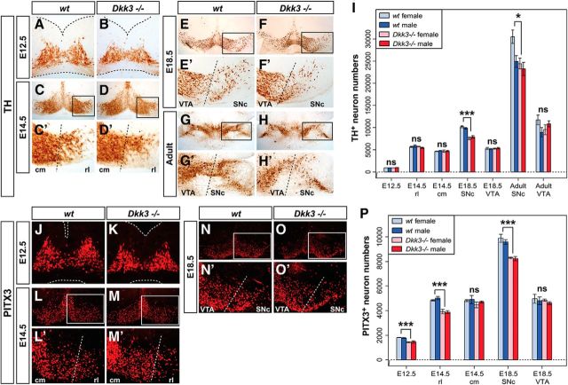
Midgestational PITX3+ cell loss and late gestational TH+ cell reduction in the Dkk3−/− midbrain. (A–H′, J–O′) Representative close-up views of the VM on coronal (A–F′, J–O′; dorsal top) and horizontal (G–H′; rostral top) sections from WT (A, C, E, G, J, L, N) and Dkk3−/− (B, D, F, H, K, M, O) embryos at E12.5 (A, B, J, K), E14.5 (C–D′, L–M′) and E18.5 (E–F′, N–O′) or adult brains (G–H′), immunostained for TH (A–H′) or PITX3 (J–O′). C′, D′, E′, F′, G′, H′, L′, M′, N′, and O′ are higher magnifications of the boxed areas in C, D, E, F, G, H, L, M, N, and O, respectively. Broken black or white lines in A, B and J, K outline the neuroepithelium and in C′, D′, E′, F′, G′, H′, L′, M′, N′, and O′ delimit the rostrolateral (rl) or SNc domain from the caudomedial (cm) or VTA domain, as indicated. I, P, Unbiased stereological quantification of TH+ (I) or PITX3+ (P) cells in female or male WT and Dkk3−/− embryos and adult brains. *p < 0.05; ***p < 0.001; ns, not significant in the Welsh t test for unequal variances.

