Figure 8.
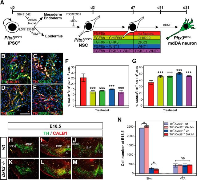
WNT/β-catenin factor treatment promotes a rostrolateral (SNc) and suppresses a caudomedial (VTA) DA neuronal fate in differentiating PSCs in vitro. A, Scheme of the monolayer protocol for the differentiation of Pitx3GFP/+ iPSCtfs into mdDA neurons. B–E, Representative confocal close-up views of TH+ (green)/CALB1+ (red) (B), TH+ (red)/KCND3+ (green) (C), TH+ (red)/DAT+ (green) (D), and TH+ (red)/GIRK2+ (green) (E) cells after treatment of differentiating Pitx3GFP/+ iPSCtfs with DKK3 + WNT1. F, G, Quantification of the proportions of CALB1+/TH+ (F) or KCND3+/TH+ (G) double-positive cells among all TH+ cells after treatment of differentiating Pitx3GFP/+ iPSCtfs with factor combinations as color coded in the scheme in A (n = 3 experiments/condition). Statistical testing for significance in F and G was done in relation to the untreated (only FGF8b-treated) cells. H–M, Representative close-up views of the VM on coronal sections (dorsal top) from WT (H–J) and Dkk3−/− (K–M) embryos at E18.5, immunostained for TH (green) and CALB1 (red). Sections were taken from rostral (H, K), intermediate (I, L), and caudal (J, M) levels of the VM. N, Quantification of the amount of TH+/CALB1− single-positive and TH+/CALB1+ double-positive cells in the SNc and VTA of E18.5 WT or Dkk3−/− embryos (n = 3 embryos/genotype). *p < 0.05 by the t test; ***p < 0.001 by one-way ANOVA relative to the FGF8b-treated cells; ns, not significant in the t test. CLi, Caudal linear nucleus; IF, interfascicular nucleus; PN, paranigral nucleus; RrF, retrorubral field; SNcd, Substantia nigra pars compacta dorsalis. Scale bar in D, 50 μm.

