Figure 1.

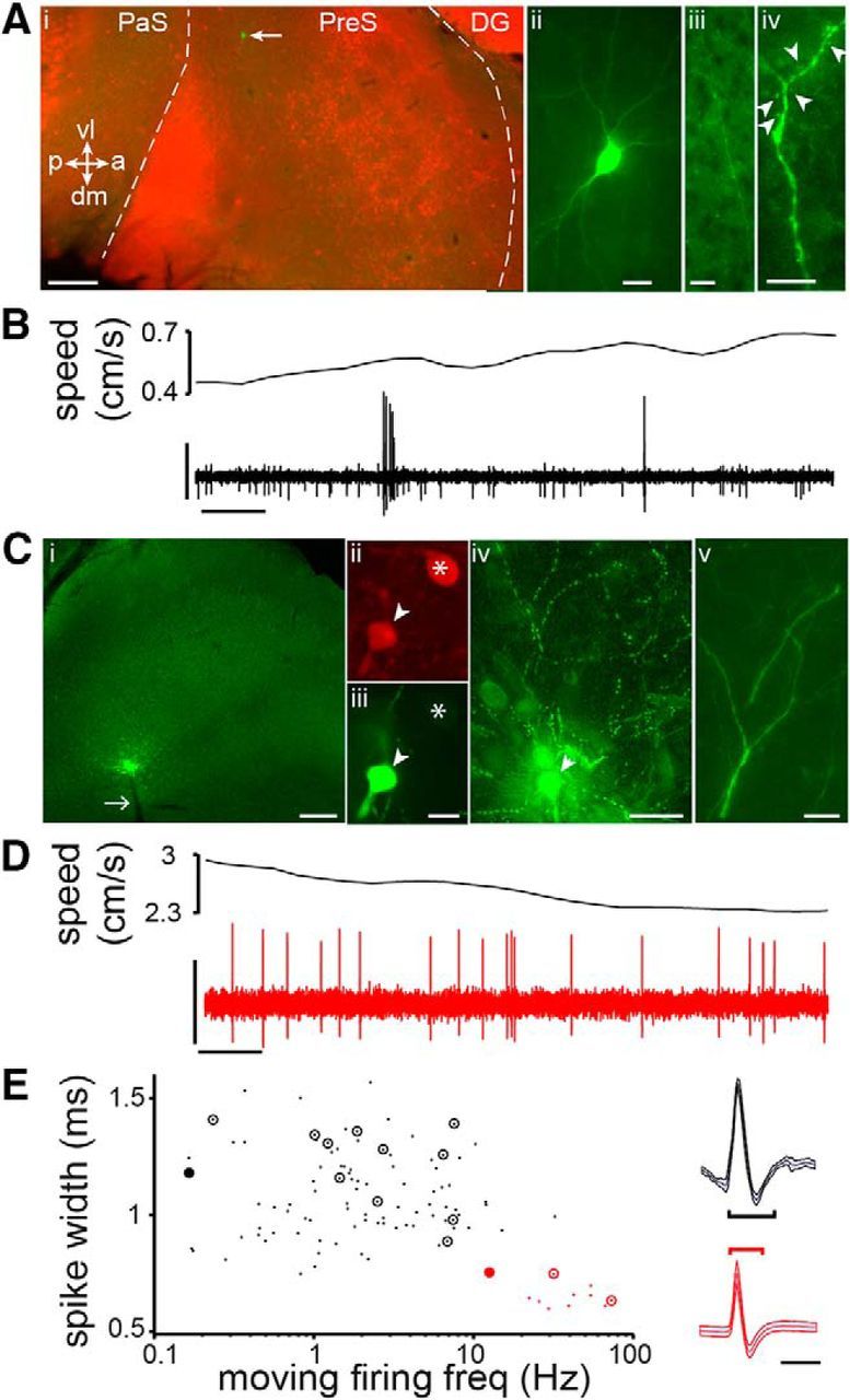
Classification of RS and FS cells in the presubiculum. A, Recovered RS neuron (green, arrow) in the superficial layers of presubiculum (i), demarcated (dashed lines) by calbindin staining (red) in this tangentially cut section (orientation: vl, ventrolateral; dm, dorsomedial; p, posterior; a, anterior). Note typical pyramidal morphology (ii), projecting axon (iii), and spiny dendrites (iv, arrowheads indicate spines). PaS, Parasubiculum; PreS, presubiculum; DG, dentate gyrus. B, Speed of the animal (top trace) and APs (bottom trace, high-pass filtered) of the neuron shown in A. C, Recovered FS neuron (green site) in the deep layers of the presubiculum (i; parasagittal section; white arrow, electrode track), immunopositive for parvalbumin (ii, iii, arrowhead, soma from labeled cell; *nearby parvalbumin-positive soma), with dense local axon arborization (iv) and nonspiny dendrites (v). D, As in B, note higher firing rate. E, FS cells (red; n = 12) and RS cells (black; n = 93) classified based on spike width (on right, mean ± SD from example cells in A–D, with measured spike widths indicated) and firing rate during movement (>2 cm/s). Identified cells, circled; filled circles, example cells from A–D. Scale bars: Ai, 0.2 mm, Aii–Aiii, 20 μm, Aiv, 10 μm; B, 2 mV, 0.1 s; Ci, 0.2 mm, Cii–Ciii, 10 μm, Civ, 50 μm, Cv, 10 μm; D, 1 mV, 0.1 s; E, 1 ms.
