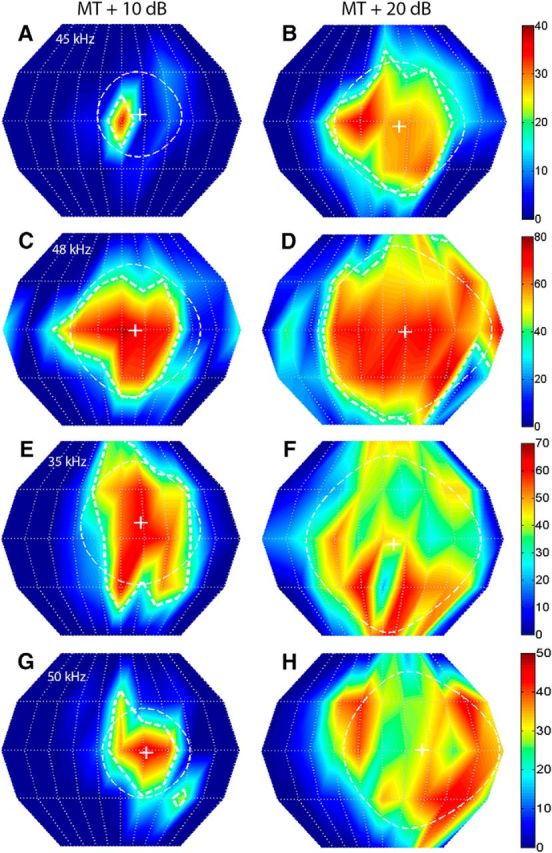
Figure 3.

The SRF properties of FMSR neurons were strongly sound level dependent. The left and right columns correspond to SRFs recorded from the same FMSR neurons, but at MT + 10 dB and MT + 20 dB, respectively. The 50% SRF contours are not shown for neurons in E–H because of the fragmenting of the SRF into multiple peaks at the higher SPL. A, B, Neuron #96A01; CF = 45 kHz; centroid azimuth = 11°, 9°; centroid elevation = 4°, −3°; gyradius = 26°, 40°. C, D, Neuron #77A02; CF = 48 kHz; centroid azimuth = 9°, 14°; centroid elevation = −1°, −1°; gyradius = 41°, 54°. E, F, Neuron #36A04; CF = 35 kHz; centroid azimuth = 13°, 7°; centroid elevation = 10°, −3°; gyradius = 38°, 54°. G, H, Neuron #86B03; CF = 50 kHz; centroid azimuth = 16°, 24°; centroid elevation = −2°, 1°; gyradius = 28°, 48°.
