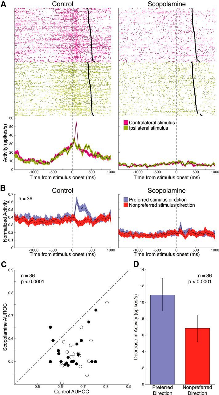
Figure 7.

Effect of scopolamine on selectivity for peripheral stimulus direction in the stimulus epoch. A, Single-neuron spike rasters and smoothed spike density functions of contralateral (pink) and ipsilateral (green) peripheral stimulus trials, for both control and 15 nA scopolamine conditions. Stimulus epoch begins 70 ms after stimulus onset and ends 120 ms after saccade onset (black diamonds). B, Normalized population spike density functions of preferred and nonpreferred stimulus direction activity in control and scopolamine conditions for 36 significantly visual stimulus-selective neurons. Scopolamine decreased both FR for preferred and nonpreferred stimulus direction and stimulus direction discriminability in the stimulus epoch. C, Scatter plot of control AUROC values (open circles represent Monkey O; filled circles represent Monkey T) compared with AUROC values in the scopolamine condition. AUROC values after scopolamine application were below the equality line, indicating reduction in direction selectivity. Population AUROC values were significantly reduced by scopolamine. D, Scopolamine elicited a stronger decrease in population FR for the preferred stimulus direction in the stimulus epoch, compared with the nonpreferred stimulus direction. Error bars indicate SEM. Significance was determined by Wilcoxon signed rank test.
