Fig. 3.
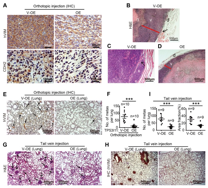
TP53I11 overexpression reduces MDA-MB-231 cells metastasis in vivo. Orthotopic injection: 231-P11-OE (OE) and vector control 231-V-OE (V-OE) cells were injected into the mammary fat pads of immunodeficient mice. Mice were euthanized 48 days later. (A) The tumors were sectioned and stained for hVIM and CDH2, and with hematoxylin and eosin (H&E). The necrotic center “n” is surrounded by a section of viable tumor rim “v” with an outer dermal layer “d” (B-D). TP53I11 overexpression reduced tumor invasion into nearby tissues. Black arrows point to the invasion locations. Red lines show the thickness of the viable tumor rim. Scale bar = 500 μm. (E) IHC staining for hVIM in lung tissue sections and (F) hVIM-positive nodules in the lungs of mice with orthotopic injections. Tail vein injection: 231-P11-OE (OE) and vector control 231-V-OE (V-OE) cells were injected into immunodeficient mice through the tail veins and the mice were sacrificed 35 days later. (G) H&E and (H) IHC (hVIM) staining of lung tissue sections. (I) The number and size of metastatic nodules. V-OE, vector control for overexpression; OE, overexpression. ***P < 0.005. P values were determined using a two-tailed student’s t-test. Results are expressed as mean ± SD from the indicated number of mice.

