
Figure 2.
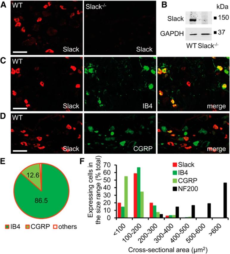
Expression of Slack channels in DRG neurons. A, Immunofluorescence of Slack in lumbar DRGs of WT and Slack−/− mice revealed specific Slack expression in 31.6 ± 1.7% (2571 cells counted, n = 3 mice) of DRG neurons. B, Western blot analysis of Slack (140 kDa) in DRG homogenates of WT and Slack−/− mice confirms the specificity of the anti-Slack antibody. GAPDH (36 kDa) was used as loading control. C, D, Typical examples of Slack immunoreactivity in nociceptor subpopulations positive for IB4 and CGRP. E, Quantitative summary of DRG neuron populations expressing Slack. Most Slack-positive cells bind IB4 (86.5 ± 1.6%, 2108 cells counted, n = 3 mice) and are therefore nonpeptidergic, whereas a few Slack-positive cells colocalize with CGRP in peptidergic DRG neurons (12.6 ± 0.33%, 1114 cells counted, n = 3 mice). F, Size distribution of Slack-positive DRG neurons compared with those expressing IB4, CGRP, and neurofilament 200 (NF200), a marker of large myelinated DRG neurons. The data demonstrate that Slack channels are nearly exclusively expressed in nociceptive DRG neurons of small and medium diameter. Scale bars, 50 μm.