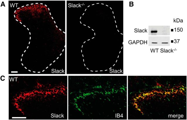
Figure 3.
Expression of Slack channels in the spinal cord. A, Immunofluorescence of Slack in the lumbar spinal cord of WT and Slack−/− mice reveals specific Slack expression in the superficial dorsal horn. B, Western blot analysis of Slack (140 kDa) in spinal cord homogenates of WT and Slack−/− mice confirms the specificity of the anti-Slack antibody. GAPDH (36 kDa) was used as loading control. C, Colocalization of Slack with IB4 shows that Slack channels are present in central terminals of sensory neurons entering the superficial dorsal horn. Scale bars, 100 μm.

