
Figure 4.
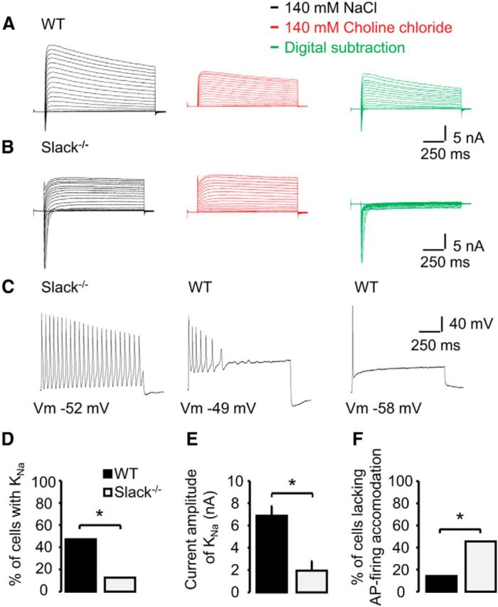
Slack generates KNa and regulates AP firing in sensory neurons. A, B, Representative current traces from whole-cell voltage-clamp recordings on IB4-positive WT (n = 17) and Slack−/− (n = 16) DRG neurons monitoring outward K+ currents in presence of 140 mm NaCl (black traces) and after replacement of NaCl with 140 mm choline chloride (red traces). Currents were elicited by protocols consisting of 1000-ms-long test pulses ranging from −80 to +110 mV in steps of 10 mV. Holding potential was −70 mV. All neurons were first recorded in presence of NaCl. To visualize the KNa current, the current traces obtained with 140 mm NaCl and 140 mm choline chloride were digitally subtracted and depicted as green traces. Please note that the absence of extracellular Na+ was verified by the lack of Na+ inward currents (red traces). C, Representative current traces from whole-cell current-clamp recordings on IB4-positive WT (n = 21) and Slack−/− (n = 22) DRG neurons. APs were elicited by 1000 ms current injections corresponding to 2–3 times the threshold of a single AP. Pictured are three observed patterns of AP firing during the current injection, i.e., no accommodation (left), accommodation (middle), and strong accommodation without repetitive firing (right). D, Percentage of DRG neurons from WT and Slack−/− mice generating outward K+ currents (KNa). E, Mean KNa current amplitudes in DRG neurons from WT (n = 8) and Slack−/− (n = 2) mice. F, Percentage of DRG neurons from WT and Slack−/− mice generating APs without accommodation, i.e., cells with repetitive firing of APs. *p < 0.05.