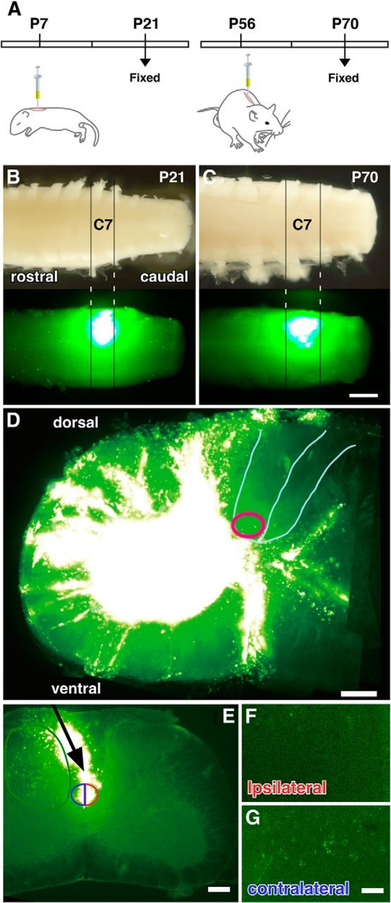
Figure 1.

Accuracy of labeling of neurons making synaptic contacts within the spinal gray matter. A, Schematic diagram of a single-injection experiment with juvenile (P7) and adult (P56) mice. Both animals were fixed 2 weeks after injection. B, C, Representative longitudinal images of juvenile (P21, B) and adult spinal cords (P70, C) showing the region where the fluorescent microspheres were injected. The spread of the microspheres was restricted to the area between the C7 and C8 nerve roots in both juveniles and adults. D, Representative transverse image of C7 at the location the microspheres were injected. Microspheres spread widely in the gray matter, but not into the ventral-most part of the dorsal column (red circle). E, F, Microsphere injection adjacent to the dorsal column in C7. A glass pipette was inserted into the vicinity of the dorsal column (E, arrow), and microspheres were spread into both sides of dorsal column (E, blue and red semicircles). No labeled cells were observed in the cortex ipsilateral to the pipette insertion (F). A small number of cells were labeled in the cerebral cortex contralateral to the side of micropipette-tip position (G), probably because microspheres were taken up by terminals distributed in the gray matter near the dorsal column, where the beads were injected (E, green circle). Scale bars: C, 500 μm; D, E, 200 μm; G, 100 μm.
