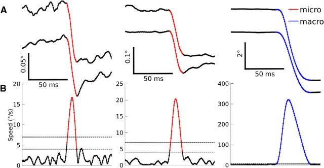
Figure 1.
Saccade detection. A, Horizontal eye position (top) vertical eye position (bottom) for three example saccades. B, Eye speed with the lenient (thin dotted line) and stringent (thick dotted line) speed thresholds overlaid (see Materials and Methods). Detected microsaccades are shown in red and macrosaccades are shown in blue.

