Figure 7.
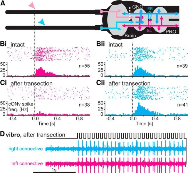
The cONv collateral in the contralateral brain hemisphere is an output branch. cONv receives input exclusively in the GNG. A, Schematic of the antennae, brain, GNG, and prothoracic ganglion (PRO), with the right (cyan) and left (magenta) cONv as well as the left (cyan) and right (magenta) afferent antennal pathways. Circles represent the sites of neck-connective recordings. ER, Electrode recording right connective; EL, electrode recording left connective. White dotted line indicates the site of transection of the right circumoesophageal connective. All data are from one continuous experiment. B, Side specificity of cONv responses to antennal stimulation and site of antennal input. Bi, Spike trains and PSTH of the left cONv during SP joint movement of the right antenna (magenta arrowhead in A, vertical dotted line). Both antennae were manually moved about the SP joint, using a fine paintbrush. Bii, Response of the right cONv to SP joint movement of the left antenna (A, cyan arrowhead). In both cases, antennal movement elicited a strong response in the contralateral cONv in the intact animal. Ci, Cii, Response of both cONvs to contralateral antennal movement after transection of the right circumoesophageal connective (A, white dotted line). The antennal response of the left, intact cONv was diminished (Ci), whereas the response of the right cONv, where the GNG-brain connection was transected, remained unaffected (Cii). D, Vibration stimulus (black), and right (cyan) and left (magenta) neck-connective recordings of cONv responses after transection of the right circumoesophageal connective (A, white dotted line). The vibration response of both cONvs remained unaffected.

