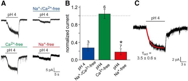
Figure 6.
Biophysical properties of proton-mediated signals. A, Representative original traces of acid-evoked whole-cell currents recorded under control conditions versus in a modified ionic environment. Extracellular Na+ and/or Ca2+ was replaced by the largely impermeable cation NMDG+ (preincubation, ≥120 s). B, Bar chart (mean ± SEM) quantitatively summarizing the results exemplified in A. Data are normalized to current densities recorded from the same neurons before cation exchange. The dashed line denotes 100%. Numbers of experiments are indicated above individual bars. The asterisk denotes statistical significance; p = 0.003 (paired-sample t test). While selective replacement of Ca2+ did not change pH-dependent currents (109 ± 9%), substitution of Na+, either alone (19 ± 6%) or together with Ca2+ (28 ± 7%), strongly reduced acid-evoked signals. C, Original recording illustrating the time course of acid-evoked currents. Activation kinetics are well fit by a single exponential function (red curve).

