Figure 1.

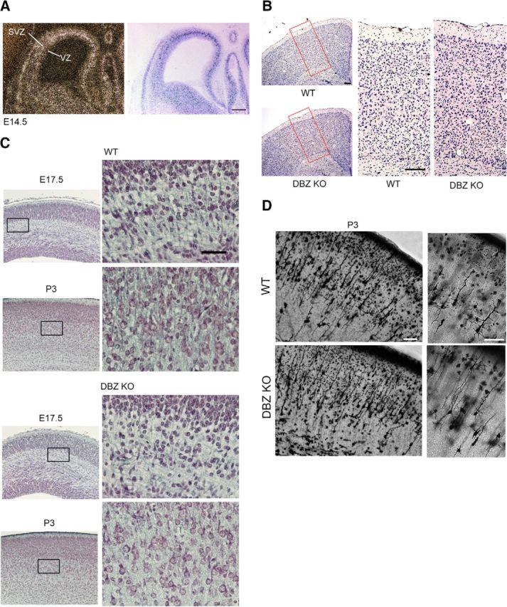
Neurite development is impaired by DBZ deletion. A, DBZ mRNA was localized primarily in the subventricular/intermediate zone (SVZ). A representative coronal section of an E14.5 cortex is shown (left). The section was counterstained with Nissl staining (right). No obvious expression was observed in the ventricular zone (VZ), whereas strong signals were observed in the SVZ. Scale bar, 200 μm. B, Histological examination of the cortices of the DBZ−/− (DBZ KO) and the littermate DBZ+/+ mice (WT). High-magnification views of the red boxes are shown on the right. Scale bars, 200 μm. C, Representative photomicrographs show coronal sections of the cortices at E17.5 and at P3 stained using Bodian's method. The DBZ−/− mice showed poor neurite development. High-magnification images in the squares on the left are shown in the next right panels. Scale bars, 100 μm. D, Examples of Golgi-impregnated P3 cerebral cortices of a WT and a DBZ KO mouse are shown. Magnified images are shown in the right. Pyramidal neurons with thin and less-branched dendrites were frequently observed in the DBZ KO. Scale bars, 40 μm.
