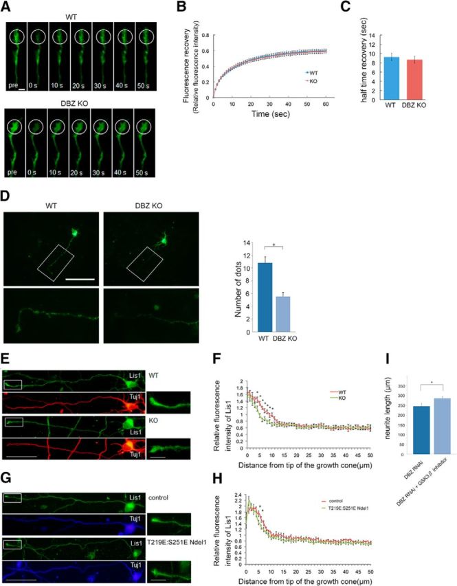
Figure 10.

Deleting DBZ or overexpressing a phosphorylation mimetic form of Ndel1 impairs the anterograde transport of DISC1 and Lis1 to the proximal ends. A, FRAP analysis of DBZ+/+ and DBZ−/− cortical neurons transfected with the mDISC1-EGFP expression vector. The indicated areas (white circles) of a growth cone were bleached, and fluorescence recovery was monitored every 1 s for 1 min. B, Quantitative fluorescence recovery data were calculated from the fluorescence intensities of the bleached areas of the DBZ+/+ and DBZ−/− cortical neurons over a period of 1 min following photobleaching. The data were normalized such that the fluorescence intensity of the prebleach sample was set to one, and the initial postbleach sample was set to zero. C, The half-time of recovery was calculated from each recovery curve. Scale bar, 5 μm. D, DISC1-HA-expressing vectors were transfected into DBZ+/+ neurons and DBZ−/− neurons. Neurons were stained with the anti-HA antibody. Magnified images of the white boxes are shown in the bottom. The occurrences of HA signals within 100 μm of the tip of axon were counted. The number of HA signals significantly decreased in the DBZ−/− neurons. Scale bar, 100 μm. E, Cortical neurons cultured from DBZ+/+ or DBZ−/− mouse embryos (E16.5) were stained with antibodies against Lis1 (green) and βIII-tubulin (Tuj1; red) at DIV3. Magnified images of the white boxes are shown on the right. F, Relative immunofluorescence intensities of Lis1 over Tuj1 within 50 μm of the tip of the growth cone were plotted. The fluorescence intensity of Lis1 decreased in the proximal side of the growth cones of the DBZ−/− neurons. G, The control empty vector or T219E:S251E Ndel1 expression vector, together with the tdTomato expression vector, were transfected at E14.5. At E16.5, tdTomato-positive neurons were collected from the cortices and dissociated for primary culture. Cortical neurons were stained with antibodies against Lis1 (green) and βIII-tubulin (Tuj1; blue) at DIV3. Magnified images of the white boxes are shown in the right. H, Relative immunofluorescence intensities of Lis1 over Tuj1 within 50 μm of the tip of the growth cone were plotted. The fluorescence intensity of Lis1 decreased in the proximal side of the growth cones in T219E:S251E Ndel1-expressing neurons. Scale bars, 50 μm (left) and 10 μm (right). I, The DBZ RNAi vector and the EGFP expression vector were transfected into the lateral ventricle at E14.5, and cortical neurons were collected and dissociated for primary culture at E16.5. Ninety-six hours after the beginning of primary culturing, the lengths of the primary neurites were measured. A GSK3β inhibitor rescued the poor neurite extension caused by acute DBZ insufficiency. The values represent the mean ± SEM; *p < 0.05.
