Figure 5.
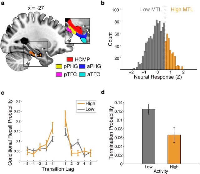
Cortical regions in the left MTL jointly support the temporal reinstatement and retrieval success hypotheses. a, A deviance map indicates a single cluster of voxels where the joint model is significantly better than the neurally naive baseline model, in a functionally defined region of interest, outlined in black (voxels identified as informative to both temporal reinstatement and retrieval success). Inset, A priori anatomical regions of interest. HCMP, Hippocampus; aPHG, anterior PHG; pPHG, posterior PHG; TFC, temporal fusisform cortex; aTFC, anterior TFC; pTFC, posterior TFC. The x-coordinates are given in MNI space. b, A histogram depicting the distribution of MTL neural responses across all recall events. Neural response is z-score normalized within a trial and is averaged across the voxels in the identified cluster. Responses >0.5 SDs above the mean response (across all trials) are labeled as high-activity recall events. c, Recall events were partitioned according to the level of activation in the cluster; periods of high activity were associated with increased temporal organization. d, Periods of high neural activity were also associated with a decreased likelihood of recall termination.

