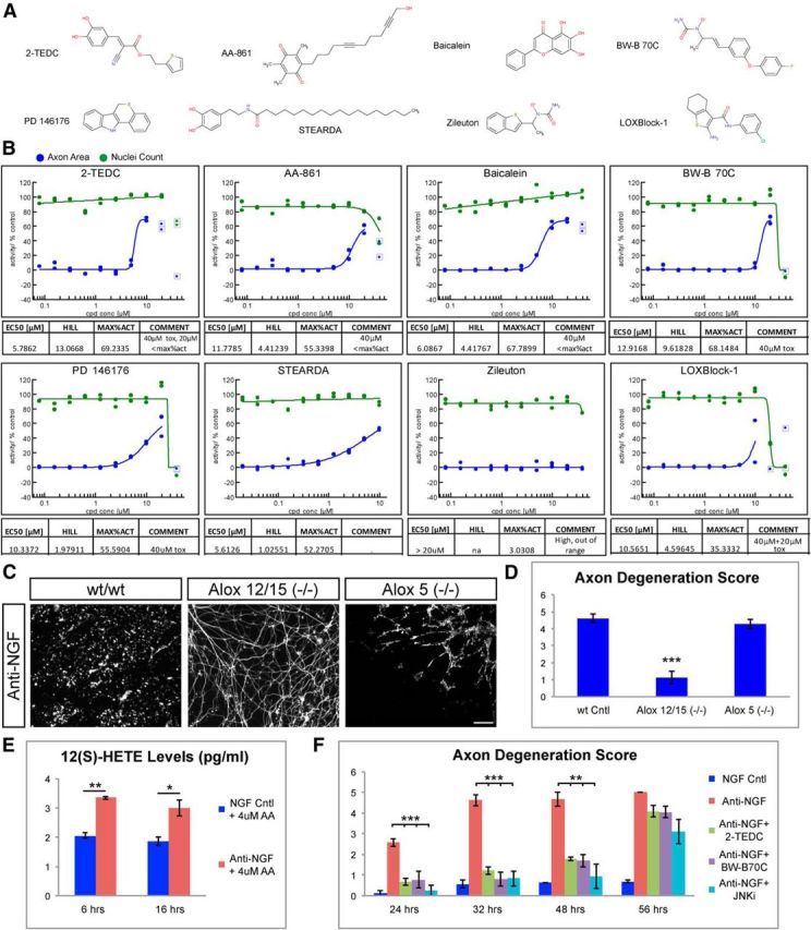
Figure 4.

Lipoxygenases are required for axon degeneration. A, Structures of lipoxygenase inhibitors tested in the assay. 2-TEDC and baicalein were hits identified in the primary screen, while the other structurally distinct lipoxygenase inhibitors were selected for follow-up studies based on published data. B, EC50 profiles of tested lipoxygenase inhibitors are shown. Graphs are representative of two to six test replicates. Compounds were tested in 10 point 1:2 dilution series with 10 μm, 20 μm[scap], or 40 μm top concentration. All tested lipoxygenase inhibitors except Zileuton showed some degree of protection. Potencies ranged between ∼6 (2-TEDC) and ∼13 μm (BW-B 70C). C, Mouse DRG neurons cultured from Alox5(−/−) and Alox12/15(−/−) mice. NGF withdrawal resulted in robust axon degeneration and loss of axons (Tuj1 staining, white) in control and Alox5(−/−) but not in Alox12/15(−/−). Scale bar, 100 μm. D, Quantification of axonal degeneration observed in C. WT = 4.63 ± 0.26, Alox12/15(−/−) = 1.23 ± 0.37, Alox5(−/−) = 4.29 ± 0.27, N = 3 independent experiments, ***p < 0.001(Student's t test, error bars indicate SEM). E, Quantification of 12(S)-HETE levels in DRG-astrocyte cocultures either 6 h or 16 h following NGF withdrawal (Anti-NGF), and in controls grown in presence of NGF (NGF Cntl). AA (4 μm) was added to the media to have an excess of lipoxygenase substrate. NGF withdrawal resulted in increased 12(S)-HETE at both time points. 6 h: NGF = 2.05 ± 0.09, anti-NGF = 3.34 ± 0.05; 16 h: NGF = 1.864 ± 0.14, anti-NGF = 3.0 ± 0.27 pg/ml. Data are representative of two independent experiments (N = 4), **p < 0.01, *p < 0.05 (Student's t test, error bars indicate SEM). F, Quantification of NGF withdrawal-induced axon degeneration at various time points following treatment with lipoxygenase inhibitors (2-TEDC or BW-B70C) or the JNK inhibitor AS601245 (JNKi). Both lipoxygenase and JNK inhibitors protect axons against degeneration up to 48 h but not 56 h post-NGF withdrawal. Relative axon degeneration score: 24 h: NGF = 0.12 ± 0.12, anti-NGF = 2.58 ± 0.19, anti-NGF + 2-TEDC = 0.66 ± 0.16, anti-NGF + BW-B 70C = 0.77 ± 0.39, anti-NGF + JNK inhibitor = 0.25 ± 0.25; 32 h: NGF = 0.56 ± 0.18, anti-NGF = 4.61 ± 0.26, anti-NGF + 2-TEDC = 1.22 ± 0.18, anti-NGF + BW-B 70C = 0.80 ± 0.35, anti-NGF + JNK inhibitor = 0.83 ± 0.37; 48 h: NGF = 0.6 ± 0, anti-NGF = 4.66 ± 0.33, anti-NGF + 2-TEDC = 1.77 ± 0.07, anti-NGF + BW-B 70C = 1.69 ± 0.29, anti-NGF + JNK inhibitor = 0.94 ± 0.59; 56 h: NGF = 0.68 ± 0.06, anti-NGF = 5 ± 0, anti-NGF + 2-TEDC = 4.08 ± 0.27, anti-NGF + BW-B 70C = 4.05 ± 0.29, anti-NGF+JNK inhibitor = 3.11 ± 0.59; ***p < 0.001, **p < 0.01 (Student's t test, error bars indicate SEM; N = 3 independent experiments, where >3 explants were scored).
