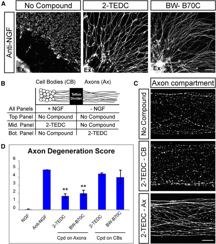
Figure 5.

Lipoxygenases act locally within axons to regulate axon degeneration. A, DRG explants (Ex) cultured in the absence of astrocytes and stained with Tuj1 to visualize axons. Axons are protected following NGF withdrawal in the presence of lipoxygenase inhibitors (2-TEDC, 18 μm; BW-B 70C, 30 μm; n = 5). Scale bar, 100 μm. B, Schematic of experiments using compartmentalized culture chambers shown in C and D in which anti-NGF is added to distal axons only and lipoxygenase inhibitor is added to either the cell body (CB) compartment or the distal axonal (Ax) compartment. C, DRG neurons grown in a compartmentalized chamber and stained with Tuj1. Axons in the distal axonal compartment are largely degenerated 28 h after NGF withdrawal (top). Addition of 2-TEDC to the cell body compartment does not protect axons from degeneration (middle), but addition to the distal axon compartment results in protection of axons (bottom). Scale bar, 50 μm. D, Quantification of axon degeneration in compartmentalized chamber treated with lipoxygenase inhibitors. NGF = 0.05 ± 0.07, anti-NGF = 4.9 ± 0, 2-TEDC on cell bodies = 4.4 ± 0.14, 2-TEDC on axons = 1.85 ± 0.07, BW-B 70C on cell bodies = 4 ± 0.84, BW-B 70C on axons = 2.35 ± 0.07; **p = 0.01, N = 2 independent experiments with multiple cultures in each experiment (Student's t test, error bars indicate SEM). Cpd, Compound.
