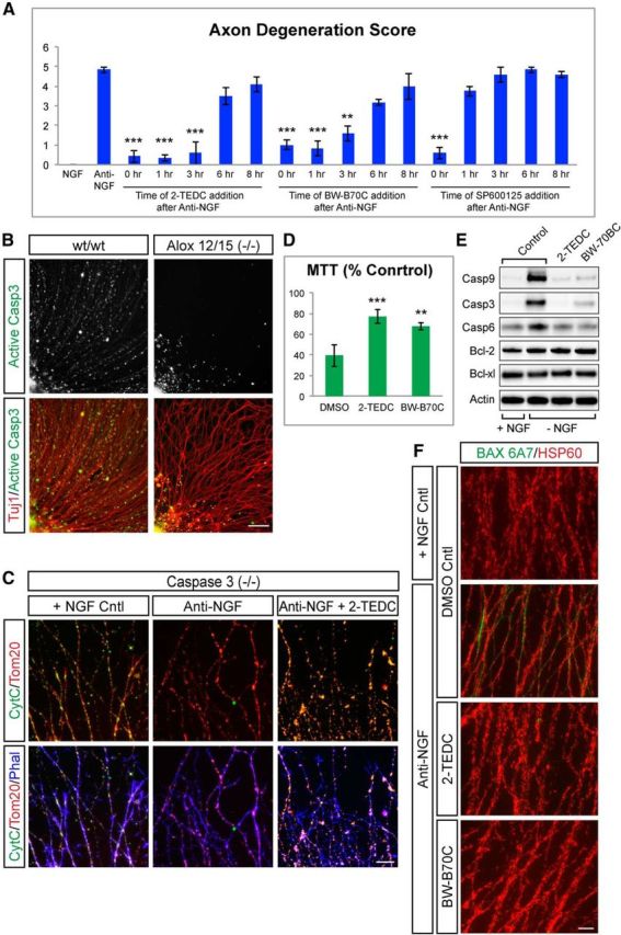
Figure 7.

Lipoxygenases are required for release of cytochrome c from mitochondria and activation of caspase-3 in axons. A, Quantification of axon protection achieved through addition of lipoxygenase inhibitor added at the times indicated following NGF withdrawal. Lipoxygenase inhibitors can be added as late as 6 h after NGF withdrawal and still protect axons from degeneration, while the JNK inhibitor (SP600125) must be added at the time of NGF withdrawal. (Anti-NGF = 4.83 ± 0.14; 2-TEDC: 0 h = 0.41 ± 0.28, 1 h = 0.33 ± 0.14, 3 h = 0.58 ± 0.57, 6 h = 3.5 ± 0.43, 8 h = 4.08 ± 0.38; BW-B 70C: 0 h = 1 ± 0.25, 1 h = 0.83 ± 0.38, 3 h = 1.58 ± 0.38, 6 h = 3.16 ± 0.14, 8 h = 4 ± 0.66; JNK inhibitor: 0 h = 0.58 ± 0.28, 1 h = 3.75 ± 0.25, 3 h = 4.58 ± 0.38, 6 h = 4.83 ± 0.14, 8 h = 4.58 ± 0.14; ***p < 0.001, **p < 0.01, *p < 0.05 (Student's t test, error bars indicate SEM; N = 3 independent experiments). B, DRG explants from wt and Alox12/15(−/−) stained for activated caspase-3 (green) and Tuj1 (red) 8 h after NGF withdrawal. Caspase-3 is activated in many wt axons, but staining is greatly reduced in Alox12/15(−/−) axons. Data are representative of four independent experiments. Scale bar, 100 μm. C, Caspase-3(−/−) DRG explants stained with cytochrome c (green), Tom20 (red), and phalloidin (blue). In the presence of NGF (left), Tom20 and cytochrome c colocalize within axonal mitochondria. Eight hours following NGF withdrawal in untreated cultures (middle), cytochrome c staining is lost, but this does not occur when cultures are treated with a lipoxygenase inhibitor (right). Data are representative of four independent experiments. Scale bar, 50 μm. D, Quantification of mitochondrial activity via an MTT assay 16 h after NGF withdrawal with or without lipoxygenase inhibitors. Mitochondrial activity is shown as percentage NGF control. MTT signal is reduced after NGF withdrawal while the addition of lipoxygenase inhibitors significantly improved mitochondrial activity. Anti-NGF = 39.45 ± 6.48%, anti-NGF + 2-TEDC = 77.15 ± 6.84%, anti-NGF + BW-B 70C = 67.62 ± 3.71%; ***p < 0.001 (Student's t test, error bars indicate SEM; N > 3 independent experiments). E, Western blots for components of the intrinsic apoptosis pathway in DRG 8 h after NGF withdrawal (− NGF) with or without lipoxygenase inhibitors compared with NGF controls (+ NGF). Levels of cleaved caspase-3, -6, and -9 increase after NGF withdrawal but are reduced in the presence of 2-TEDC or BW-B 70C. No change is observed in Bcl-xl and Bcl-2 levels in any condition. F, Immunostaining with a Bax antibody specific for the active conformation in wt DRG explants 6 h following NGF withdrawal with or without lipoxygenase inhibitors. Bax 6A7 (green), HSP60 (red). NGF withdrawal results in increased axonal 6A7 staining that does not occur when cultures are treated with 2-TEDC or BW-B 70C. Data are representative of four independent experiments. Scale bar, 50 μm.
