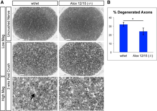
Figure 8.
Alox12/15(−/−) displays reduced axon degeneration after optic nerve crush. A, Representative images of PPD-stained proximal optic nerve sections from wt and Alox12/15(−/−) animals 3 weeks after nerve crush. Loss of intact axons can be readily observed 3 weeks after nerve crush in both genotypes compared with uncrushed contralateral nerves, though the amount of axonal degeneration in Alox12/15(−/−) mice was reduced compared with and without controls. Arrow indicates areas devoid of axons in crushed nerves. Low-magnification (Low Mag) scale bar, 50 μm; high-magnification (High Mag) scale bar, 20 μm. B, Quantification of images shown in A. wt = 32.19 ± 2.29% and Alox12/15(−/−) = 24.04 ± 4.97%; *p < 0.05 (Student's t test, error bars indicate SEM, N = 5 animals/genotype, with 10 sections quantified from each nerve).

