Figure 1.
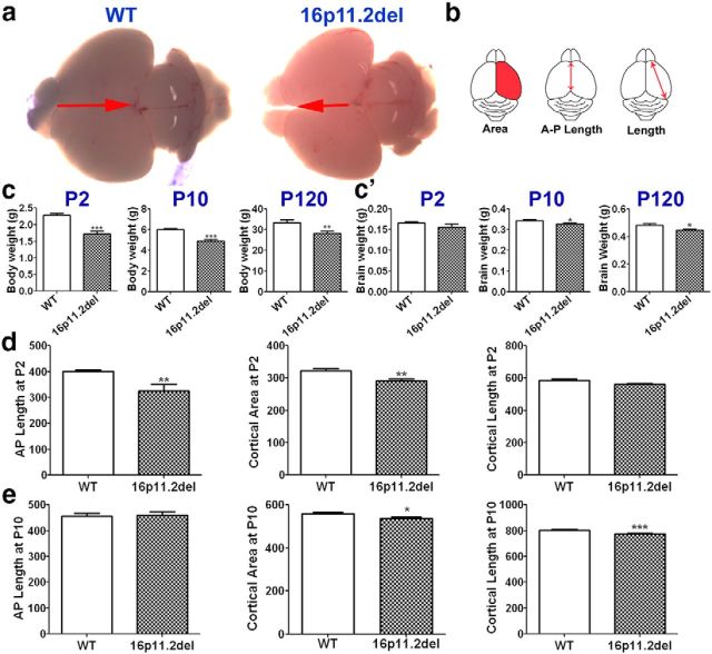
16p11.2del mice display reduced body weight, brain weight, and cortical size. a, Images of whole brains of WT (left) and 16p11.2del (right) mice at P2. c, 16p11.2del mice had reduced body weight at P2 (***p = 0.0008), P10 (***p < 0.0001), and P120 (**p = 0.0075). c′, 16p11.2del mice exhibited unchanged brain weight at P2 but decreased brain weight at P10 (*p = 0.021) and P120 (*p = 0.018). Measurements of cortical parameters (pictured in b) were obtained at P2 (d) and P10 (e). d, At P2, 16p11.2del cortices show decreased anteroposterior length (**p = 0.004), cortical length (p = 0.056), and cortical area (**p = 0.0096) compared with WT. e, At P10, 16p11.2del cortices showed decreased cortical length (**p = 0.0068) and cortical area (*p = 0.045). ndel = 5–7; nwt = 5–7 at P2, P10, and P120. *p < 0.05. **p < 0.01. ***p < 0.0001.

