
Figure 1.
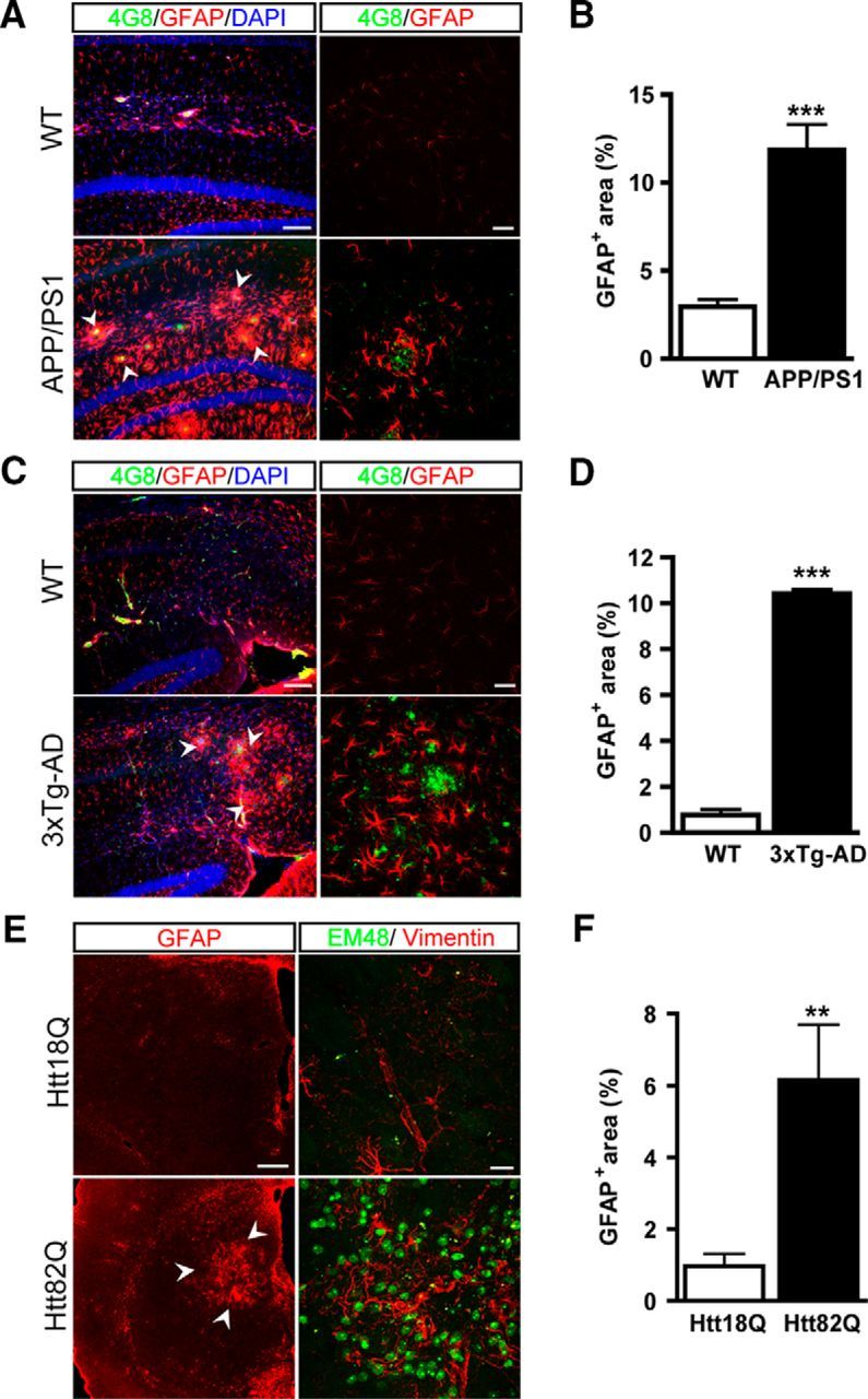
Mouse models of AD and HD display astrocyte reactivity in vulnerable regions of the brain. A–C, Images of brain sections from mouse models of AD showing double staining for amyloid plaques (4G8, green) and astrocytes (GFAP, red). A, GFAP is strongly expressed in hippocampal astrocytes of 8-month-old APP/PS1dE9 mice around amyloid depositions (arrowheads) in the stratum lacunosum moleculare and the dentate gyrus. B, Quantification of the GFAP+ area in the hippocampus of APP/PS1dE9 and WT mice. C, In the subiculum, 3xTg-AD mice display amyloid depositions that are surrounded by GFAP+ reactive astrocytes (arrowheads). D, Quantification of the GFAP+ area in the subiculum of 12-month-old 3xTg-AD mice and age-matched WT mice. E, Mice injected with lenti-Htt82Q in the striatum display EM48+ aggregates of mHtt (green). Expression of the mHtt in striatal neurons leads to astrocyte reactivity (arrowheads) as shown by increased GFAP and vimentin staining (red). F, Quantification of the GFAP+ area in the lenti-Htt82Q-injected striatum relative to the control striatum injected with lenti-Htt18Q. n = 3–6 per group. **p < 0.01, ***p < 0.001. Scale bars: A and C, left, 100 μm; right, 20 μm; E, left, 200 μm; right, 20 μm.